Skip Navigation
Skip to contents

Intest Res : Intestinal Research

IMPACT FACTOR

Articles

Page Path
HOME > Intest Res > Volume 19(1); 2021 > Article
Original Article
Miscellaneous
Patients with celiac disease are at high risk of developing metabolic syndrome and fatty liver
Ashish Agarwalorcid, Alka Singhorcid, Wajiha Mehtaborcid, Vipin Guptaorcid, Ashish Chauhanorcid, Mahendra Singh Rajputorcid, Namrata Singhorcid, Vineet Ahujaorcid, Govind K. Makharia,orcid
Intestinal Research 2021;19(1):106-114.
DOI: https://doi.org/10.5217/ir.2019.00136
Published online: April 22, 2020

Department of Gastroenterology and Human Nutrition, All India Institute of Medical Sciences, New Delhi, India

Correspondence to Govind K. Makharia, Department of Gastroenterology and Human Nutrition, All India Institute of Medical Sciences, Ansari Nagar, New Delhi 110029, India. Tel: +91-11-26546546, Fax: +91-11-26588091, E-mail: govindmakharia@gmail.com
• Received: October 22, 2019   • Revised: December 30, 2019   • Accepted: January 31, 2020

© Copyright 2021. Korean Association for the Study of Intestinal Diseases. All rights reserved.

This is an Open Access article distributed under the terms of the Creative Commons Attribution Non-Commercial License (http://creativecommons.org/licenses/by-nc/4.0/) which permits unrestricted non-commercial use, distribution, and reproduction in any medium, provided the original work is properly cited.

prev next
  • 12,336 Views
  • 225 Download
  • 40 Web of Science
  • 44 Crossref
  • 46 Scopus
  • Background/Aims
    Gluten-free diet has an excess of fats and simple sugars and puts patients with celiac disease at risk of metabolic complications including metabolic syndrome and fatty liver. We assessed prevalence of metabolic syndrome and fatty liver in two cohorts of celiac disease.
  • Methods
    Study was done in 2 groups. In group 1, 54 treatment naïve patients with celiac disease were recruited. Of them, 44 returned after 1-year of gluten-free diet and were reassessed. In group 2, 130 celiac disease patients on gluten-free diet for ≥1 year were recruited. All patients were assessed for anthropometric and metabolic parameters and fatty liver. Metabolic syndrome was defined as per consensus definition for Asian Indians. Fatty liver was defined as controlled attenuation parameter value >263 decibels by FibroScan.
  • Results
    In group 1, of 44 treatment naïve patients with celiac disease, metabolic syndrome was present in 5 patients (11.4%) at baseline and 9 (18.2%) after 1 year of gluten-free diet. Patients having fatty liver increased from 6 patients (14.3%) at baseline to 13 (29.5%) after 1year of gluten-free diet (P=0.002). In group 2, of 130 patients with celiac disease on gluten-free diet for a median duration of 4 years, 30 out of 114 (26.3%) and 30 out of 130 patients (23%) had metabolic syndrome and fatty liver, respectively.
  • Conclusions
    Patients with celiac disease are at high risk of developing metabolic syndrome and fatty liver, which increases further with gluten-free diet. These patients should be assessed for nutritional and metabolic features and counseled about balanced diet and physical activity regularly.
Celiac disease (CeD) is usually considered to lead to undernutrition and low weight both in children and adults; in recent times, however, many patients with CeD are found to be not only overweight but some even being obese [1]. In a retrospective study, we observed that 54.8% of treatment naïve patients with CeD had their body mass index (BMI) within normal limits, 8.1% of them, however, were either overweight or obese [1]. Numerous other studies have made similar observations and up to 44% of the patients were found to be overweight or obese at the time of diagnosis of CeD [2,3]. Furthermore, many studies have uniformly shown that underweight patients at the time of diagnosis tend to gain weight on gluten-free diet (GFD) [4-6]. Contradictory to that while 22% to 82% of overweight and obese patients gain weight on a GFD [4,7]; some of the overweight and obese patients with CeD lose weight on GFD [6,8]. As described above, many studies have looked into the effect of a GFD on BMI but the association between CeD and metabolic syndrome has not been explored in much detail. In a recent prospective study, Tortora et al. [9] reported an increase in the prevalence of metabolic syndrome from 2% at the time of diagnosis to 30% after 12 months of GFD. Although there is emerging evidence that a sizeable proportion of patients with CeD gain weight and end up developing metabolic syndrome, the pathogenesis of this condition is not very clear. Processed GFD is considered to be nutritionally imbalanced that lead to a higher intake of carbohydrates with a higher glycemic index [10]. Furthermore, high quantity of saturated fat used in preparing GFD processed food to make them palatable, also add to higher calorie intake [10]. Moreover, a phenomenon of hyperabsorption have been described after the restoration of intestinal mucosal function with GFD in patients with CeD [10]. A differential secretion pattern of brain-gut axis hormones has also been described in patients with both untreated and treated CeD that may underlie weight gain and development of the metabolic syndrome [10,11].
In the background of obesity and metabolic syndrome, many patients develop nonalcoholic fatty liver disease (NAFLD). Fatty liver, if untreated, can lead to cirrhosis in up to 3% patients [12]. There are studies to suggest that patients with CeD are at 3-fold higher risk of developing NAFLD [13-18]. Furthermore, patients with CeD are at a higher risk of cardiovascular diseases (hazard ratio, 1.10; 95% confidence interval [CI], 1.03–1.28) and stroke (odds ratio, 1.11; 95% CI, 1.02–1.20) [19].
Since there is a limited data globally and none from Asia on the prevalence of metabolic syndrome and fatty liver disease in patients with CeD, we conducted a study to assess the prevalence of metabolic syndrome in treatment naïve patients with CeD before and after 1 year of GFD prospectively and in another cohort of patients with CeD who were on GFD for at least 1 year.
This prospective study was conducted at the Celiac Disease Clinic, All India Institute of Medical Sciences, New Delhi. The study protocol was approved by the Institutional Ethics Committee (approval No. IECPG-527/26.10.2016) and written informed consent was taken from each participant. The study protocol conforms to the ethical guidelines of the 1975 Declaration of Helsinki as reflected in a priori approval by the institution’s human research committee.
The study was conducted in 2 groups. In group 1, treatment naïve biopsy-proven adult patients with CeD presenting to the clinic between November 2016 and October 2017 were assessed for metabolic syndrome and fatty liver at the baseline and then they were reassessed after 1 year of GFD. In group 2, we assessed patients with CeD who were already on GFD for more than 1 year for the presence of metabolic syndrome and fatty liver.
Pregnant patients, patients consuming more than 2 standard alcoholic drinks per day, smokers, patients with preexisting diagnosed chronic liver disease, patients with a co-morbid illness such as cardiac diseases, renal diseases, pulmonary diseases, uncontrolled hypothyroidism, malignancy, untreated infection, and uncontrolled type I diabetes were excluded. The diagnosis of CeD was made based on standard criteria, i.e. a positive anti-tissue transglutaminase and/or anti-endomysium antibody and presence of villous abnormality of modified Marsh grade 2 and above [20].
1. Study Parameters
All the patients in both the groups underwent assessment for the demographic and clinical parameters including ethnicity, personal or family history of prior hyperlipidemia, hypertension, diabetes, peripheral vascular disease, coronary artery disease, NAFLD or obstructive sleep apnea. They were also assessed for blood pressure (mmHg), height (cm), weight (kg), BMI (kg/m2), waist and hip circumference (as per WHO STEPwise approach to surveillance protocol) [21], and waist-hip ratio. Baseline laboratory work included complete hemogram, liver function tests, renal function tests, fasting serum glucose, fasting lipid panel and IgA anti-tissue transglutaminase antibodies. These clinical and laboratory parameters were obtained again at a follow-up of 1 year on GFD in the participants included in group 1.
2. Measurement of Liver Stiffness and Controlled Attenuation Parameter
Liver stiffness measurement and controlled attenuation parameter (CAP) measurement were performed on a FibroScan touch 502 (Echosens, Paris, France) as per the standard recommendations. All measurements were performed by a single operator and measurements were done in fasting state in morning hours. The operator of FibroScan was blinded to the clinical and biological data of the patients. FibroScan was done at baseline and after 1 year of follow-up in the participants of group 1 and at the time of recruitment in group 2.
3. Body Composition Analyses
Body composition was assessed using bioelectrical impedance analysis using a Tanita TBF-215 leg-to-leg portable impedance analyzer (Tanita, Tokyo, Japan). The body weight and impedance were measured from which the fat mass, percentage fat and fat-free mass were calculated. The fat mass was computed as the difference between body weight and fat-free mass. Fat mass index (FMI) was calculated as fat mass/height2. Height was measured using the built-in height rod in the instrument. For all patients, 0.5 kg was deducted to account for their clothes. All measurements were made empty stomach after 12 hours of overnight fast, without any intake of liquids. Three repeat measurements were recorded and the mean value was taken as final.

1) Treatment of CeD

All treatment naïve patients were counseled by an expert and dedicated dietician (W.M.) and all of them were followed at 3 months interval for reconciling and assessment of adherence. Adherence was assessed based on detailed dietary history by the dietician and was graded as follows: (1) excellent: participant ate gluten once per 6 months; (2) good: participant ate gluten 0–1 times per month; (3) poor: participant ate gluten 2–3 times per month, participant ate gluten greater than two times per week or participant does not follow GFD.
4. Definition of Outcome Measures
Consensus definition for Asian Indians [22] was used for the diagnosis of metabolic syndrome. It includes the following 5 parameters: (1) abdominal obesity measured as waist circumference (>90 cm for males and >80 cm for females); (2) hypertriglyceridemia (serum triglycerides level >150 mg/dL); (3) low levels of serum high density lipoprotein (HDL) cholesterol (<40 mg/dL for males and <50 mg/dL for females); (4) hypertension (blood pressure >130/85 mmHg); and (5) fasting hyperglycemia (fasting blood sugar [FBS] >100 mg/dL). All those patients satisfying at least 3 of 5 criteria were classified as having metabolic syndrome.

1) Fatty Liver

Patients having a CAP value of more than 263 decibels, as determined by FibroScan technique, were classified as having fatty liver. The severity of fatty liver was defined as follows: grade I: CAP 263–286.9 dB; grade II: CAP 287–295.9 dB; grade III: CAP ≥296 dB [23].

2) Obesity

BMI was calculated as weight/height2 (kg/m2) and was graded as per the BMI classification suggested by the Consensus Statement for Asian Indians as follows [22]: underweight: BMI <18.5 kg/m2; normal: BMI 18.5–22.9 kg/m2; overweight: BMI 23–24.9 kg/m2; and obese: BMI ≥25 kg/m2.
5. Statistical Analysis
Statistical analysis was performed using SPSS version 13.0 (SPSS Inc., Chicago, IL, USA). Continuous variables were expressed as mean±standard deviation or median (range) as appropriate. Categorical variables were compared by chi-square test or Fisher exact test as appropriate. Continuous variables were compared by Student t-test or Mann–Whitney test as appropriate.
1. Comparison of Anthropometric and Metabolic Parameters of Patients before and after 1 Year of GFD: Group 1
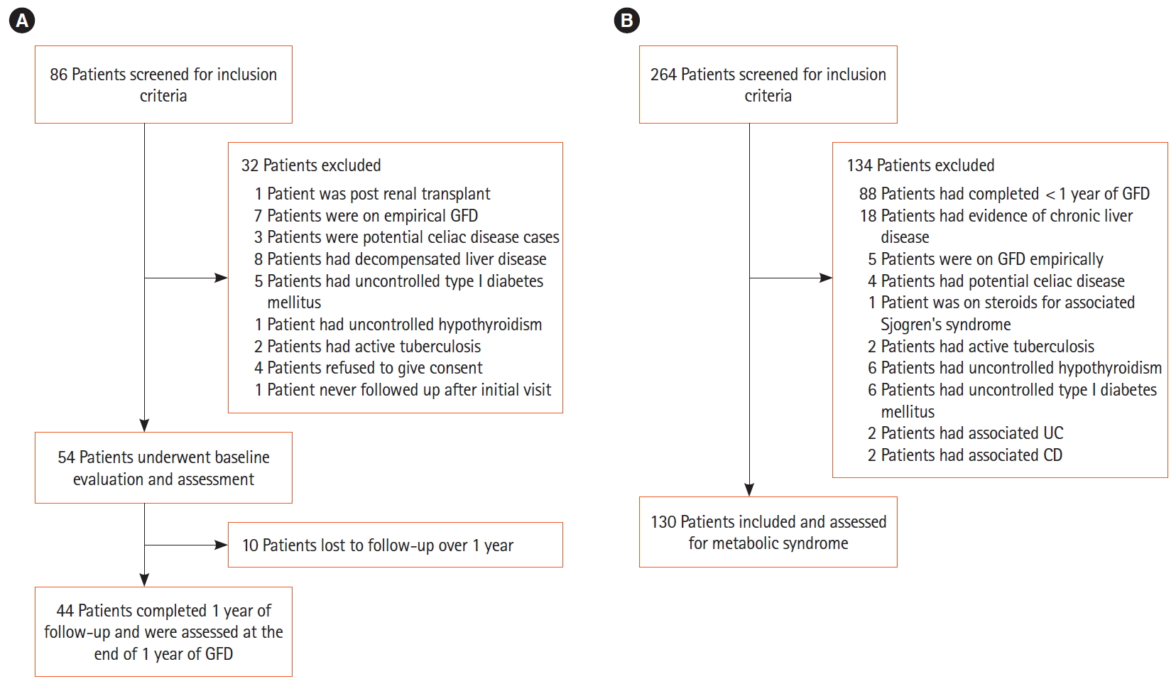
In this study, we screened 86 consecutive patients with CeD for inclusion in the study, of which 54 treatment naïve CeD patients were included and they underwent baseline assessment and were started on GFD under the care of a nutritionist. Of them, 10 patients were lost to follow-up and 44 patients were reassessed after 1 year of GFD (Fig. 1A). Of 54 treatment naïve patients, 9 patients (16.6%) were overweight or obese, 13 (24.1%) had fatty liver and 5 (11.1%) had evidence of metabolic syndrome at the baseline.

1) Changes in BMI and Obesity

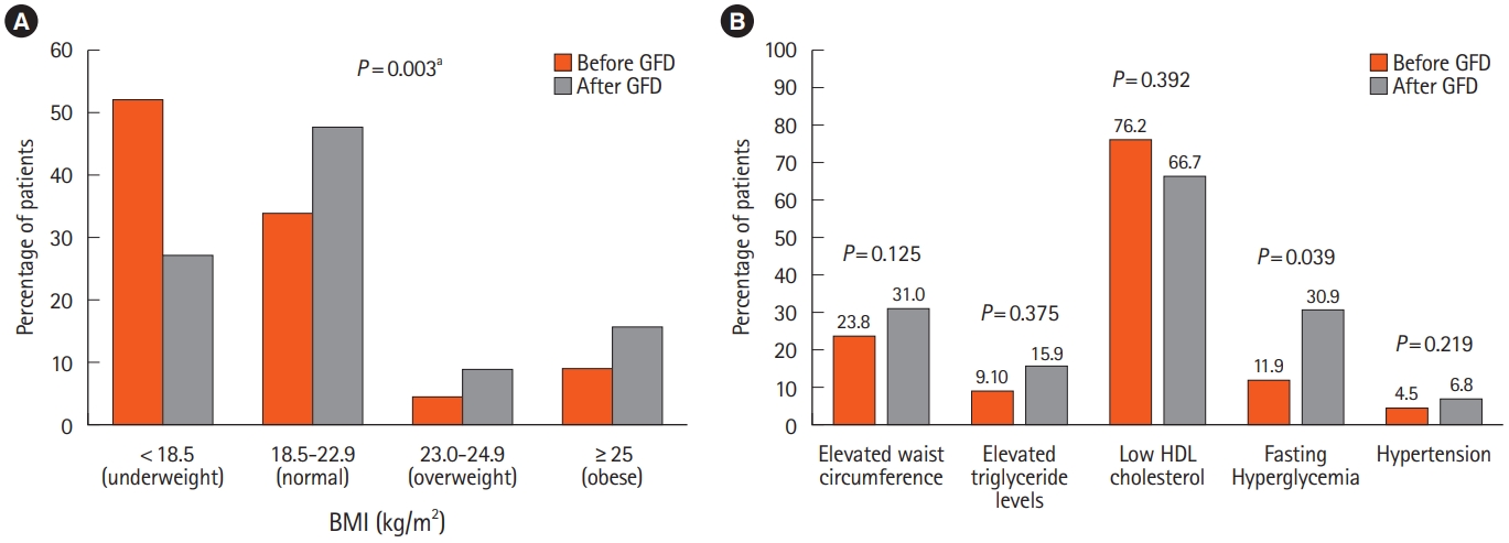
Though there was no significant increase in the height (P=0.3) of the patients, there was a significant rise in the body weight after 1 year of GFD (before 49.0±13.7 kg vs. after 53.2±13.3 kg; P<0.001) thus resulting in a significant increase in BMI (P=0.003). The proportion of overweight and obese patients increased from 13.6% at the baseline to 25% after 1 year of GFD (Fig. 2A). This was also associated with an increase in the mean waist circumference (before 76.9±10.9 cm vs. after 80.1±12.5 cm; P=0.003) and the mean hip circumference (before 84.6±7.9 cm vs. 88.0±8.7 cm after; P<0.001). On body composition analysis, in comparison to the baseline, there was a significant increase in the median body fat percentage (before 10% [interquartile range (IQR), 2.5–31] vs. after 12% [IQR, 1.8–37]; P=0.001) and FMI (before 1.8 kg/m2 [IQR, 0.44–9.1] vs. after 2.4 kg/m2 [IQR, 0.3–10.5]; P=0.003) after 1 year of GFD (Table 1).

2) Metabolic Syndrome in Treatment Naïve Patients with CeD and 1 Year after GFD

Nine of 44 patients (18.2%) had evidence of metabolic syndrome after 1 year of GFD, compared to 5 of 44 patients (11.4%) before starting GFD, suggesting a rise in the prevalence of metabolic syndrome after just 1 year of GFD (Table 2). Of the 5 patients with metabolic syndrome before initiation of GFD, 4 continued to have metabolic syndrome. One patient ceased to satisfy the criteria for metabolic syndrome, mainly because of improvement in the value of HDL, though he continued to have elevated FBS level and high waist circumference. Five new patients developed metabolic syndrome at 1 year of GFD.
Among the individual parameters of the metabolic syndrome, the number of patients having hyperglycemia increased from 5 (11.9%) to 13 (30.9%) (P=0.039) and hypertriglyceridemia from 4 (9.1%) to 7 (15.9%) (P=0.375). There was an increase in the patients with elevated waist circumference (before 22.7% vs. after 31.8%; P=0.125) and 1 patient developed hypertension after starting GFD. Though a slight decrease in the proportion of patients with low HDL levels was noted (before 74.4% vs. after 63.6%; P=0.392), the change was mainly in male patients (Fig. 2B).

3) Fatty Liver

While there was no significant difference in the liver stiffness (P=0.098), there however was a significant increase in the mean CAP values from 213.5±46.9 dB to 230.8±50.1 dB (P=0.001) after 1 year of GFD. The proportion of patients having fatty liver increased from 6 patients (14.3%) at baseline to 13 patients (29.5%) after 1 year of GFD (P<0.002). Furthermore, there was an increase in the proportion of patients having a more severe grade of fatty liver with 8 patients (18.2%) patients having grade III fatty liver after 1 year of GFD compared to 1 patient (2.4%) at baseline.
2. Anthropometric and Metabolic Parameters of Patients with CeD on GFD for More Than 1 Years: Group 2
For the retrospective study, 264 patients with CeD who were on GFD for more than 1 year were screened and of them, 130 patients who met the inclusion and exclusion criteria were included. They underwent evaluation for metabolic syndrome and fatty liver (Fig. 1B).

1) BMI and Obesity

Mean BMI was 22.5±4.3 kg/m2 with 25 patients (19.2%) being underweight, 54 patients (41.5%) with BMI within normal range and 51 patients (39%) in overweight (13.8%) and obese (25.4%) category. On body composition analyses, median body fat percentage was 18.0% (range, 1.1%–40.0%) and both body fat percentage and the FMI was elevated above the recommended cutoff in 19.2% patients (Table 1).

2) Metabolic Syndrome

Of 130 patients, 114 patients had all the 5 parameters available for the detection of metabolic syndrome (Table 2). Thirty patients (26.3%) satisfied the criteria for metabolic syndrome as per the consensus definition for Asian Indians. Of these, 51 patients (44.7%) had abdominal obesity evidenced by waist circumference above the cutoff for metabolic syndrome. Fasting hyperglycemia (FBS >100 mg/dL) was present in 40 patients (35.1%). Mean HDL was below the recommended cutoff value for MS in 58 patients (50.9%). Fasting hypertriglyceridemia (TG >150 mg/dL) was noted in 16 patients (14.0%) while hypertension was noted in 6 patients (5.3%).

3) Fatty Liver

On FibroScan, mean liver stiffness measurement was 4.9±1.2 kPa and mean CAP was 226.7±50.2 dB. Twenty-three percent of patients had fatty liver (CAP ≥263 dB) with 11.5%, 3.8%, and 7.7% patients having grade I, grade II and grade III fatty liver, respectively.
Contrary to the popular belief, the present study shows a relatively high prevalence of metabolic syndrome in treatment naïve patients with CeD. Of 44 treatment naïve patients with CeD, 5 (11.4%) had metabolic syndrome at the baseline. There was a further rise in the prevalence of metabolic syndrome with 9 of 44 patients (18.2%) having features of metabolic syndrome 1 year after the institution of GFD. Furthermore, in patients who were on GFD for more than 1 year, the prevalence of metabolic syndrome was even higher with 30 of 114 patients (26.3%) having metabolic syndrome, similar to that in the general population [24,25]. The higher prevalence of metabolic syndrome in treatment naïve patients with CeD patients in the present study compared to an earlier study by Tortora et al. (11% vs. 2%) [9], could be related to the difference in the definition of metabolic syndrome (consensus definition for Asian Indians in the present study vs. Adult Treatment Panel III criteria used by Tortora, et al.)
Furthermore, present study demonstrated a significant increase in the prevalence of fatty liver from 14.3%, as demonstrated on FibroScan, at the time of diagnosis of CeD, to 29.5% 1 year of GFD (P=0.002). The prevalence of fatty liver in those patients with CeD who were following on GFD for more than 1 year was 23%, which is similar to the population prevalence of the fatty liver disease varying from 16% to 32% in India [26-28]. The results of the present study show a higher prevalence of fatty liver than that reported in an earlier study [18]. The higher prevalence of fatty liver in these patients may partly be because of use of a more sensitive and more reliable technique such as FibroScan for detection of fatty liver in comparison to ultrasonographic examination, which is mostly qualitative, subjective and operator dependent and more likely to miss the lower degrees of fat accumulation in the liver [29].
While 13% patients had a BMI in overweight/obese category at the diagnosis of CeD, the proportion increased to approximately 25% after 1 year of GFD in them. Overall 89% of patients gained weight across different BMI categories after initiation of GFD. Interestingly, the increase in the BMI was mainly in the body fat rather than in the muscle mass, as suggested by the rise is the body fat percent and FMI in them.
The development of metabolic syndrome, obesity, and fatty liver is a slow process and all the metabolic abnormalities and their severity increase with increasing duration. The occurrence of metabolic syndrome and fatty liver in patients with CeD, which otherwise is a malabsorptive state, is an important observation and such a disease have long-term adverse consequences. It calls for our attention because it is still believed that patients with CeD are undernourished and overweight/obesity is not a concern in them. Furthermore, GFD is the only available treatment for CeD at this point of time, and a long-term GFD can lead to perpetual and progressive worsening of metabolic derangements with increasing duration of GFD and thus predisposing them to higher cardiovascular risk. Therefore, it is imperative that we recognize metabolic abnormalities as a reality in patients with CeD and hence initiate appropriate preventive strategies, including counselling for physical activities and a well-balanced GFD. It is also relevant to advocate to the health authorities to legislate and monitor the calories and fat contents of gluten-free food items.
While the strength of the present study includes inclusion of not only the patients with CeD and their follow-up at 1 year but also the inclusion of a relatively large number of patients with CeD who were already on GFD for more than 1 year. There are however few shortcomings of the present study. The follow-up of patients was only for 1 year and a longer follow-up would have highlighted the longer-term adverse consequences in these patients. While these patients were counseled by a trained CeD dietician, we could not collect systematically the daily calories and nutrition intake and energy expenditure data in them which could have helped further substantiate if GFD was directly related to the development of the metabolic derangements in this subset of patients.
In conclusion, patients with CeD are at high risk of developing metabolic syndrome and fatty liver with the initiation of GFD. Patients with CeD on GFD should be assessed for nutritional and metabolic features at regular interval. They should be advised to avoid adding excess fats and sugars to the homemade gluten-free products and avoid taking commercially available GFD with high fats and sugars like cakes, cookies, sweets and biscuits. They should be counseled about a balanced diet and physical activity.

Funding Source

The authors received no financial support for the research, authorship, and/or publication of this article.

Conflict of Interest

No potential conflict of interest relevant to this article was reported.

Author Contribution

Conceptualization: Makharia GK, Agarwal A. Methodology: Agarwal A, Makharia GK, Singh A, Mehtab W. Formal analysis: Agarwal A, Makharia GK, Chauhan A. Project administration: Makharia GK. Visualization: Makharia GK, Agarwal A. Writing - original draft: Agarwal A, Singh A, Chauhan A, Rajput MS. Writing - review and editing: Agarwal A, Makharia GK, Ahuja V, Makharia GK, Singh N, Mehtab W, Gupta V. Approval of final manuscript: all authors.

Others

We acknowledge our patients for participation in the study and the staff and residents of the Celiac Disease Clinic, All India Institute of Medical Sciences.

Fig. 1.
(A) Flowchart showing enrolment of newly diagnosed celiac disease patients and follow-up. (B) Flowchart showing enrolment of patients with celiac disease on gluten-free diet (GFD) for more than 1 year. UC, ulcerative colitis; CD, Crohn’s disease.
ir-2019-00136f1.jpg
Fig. 2.
(A) Comparison of body mass index (BMI) before and after gluten-free diet (GFD). (B) Individual criteria of metabolic syndrome before and after 1 year of GFD. aP-value for the BMI distribution before and after GFD.
ir-2019-00136f2.jpg
Table 1.
Comparison of Demographic, Clinical, Biochemical, Anthropometric and Metabolic Parameters of Patients before and after 1 Year of GFD and Patients on Long-term GFD
Characteristic Before GFD (n = 44) After GFD (n = 44) P-valuea Patients on long-term GFD (n = 130)
Sex -
 Male 18 (41.0) 18 (41.0) 53 (40.8)
 Female 26 (59.0) 26 (59.0) 77 (59.2)
Age (yr) 29.5 ± 11.3 - - 33.0 ± 13.1
Hemoglobin (g/dL) 10.4 ± 2.6 12.4 ± 1.8 < 0.001 12.1 ± 1.8
Hemoglobin categories (g/dL) < 0.001
 <8 10 (23.3) 0 2 (1.6)
 8–12 17 (39.5) 19 (43.2) 59 (45.4)
 > 12 16 (37.2) 25 (56.8) 67 (51.5)
Corpuscular volume (fL/cell) 77.2 ± 10.6 81.0 ± 7.0 0.009 83.4 ± 8.8
Corpuscular hemoglobin (pg/cell) 25.2 ± 4.3 28.2 ± 10.3 0.080 27.2 ± 2.9
Corpuscular hemoglobin concentration (g/dL) 30.8 ± 1.7 31.4 ± 2.1 0.014 31.5 ± 1.8
AST (U/L) 32.8 ± 16.0 31.7 ± 14.8 0.420 28.5 (11.0–83.0)
ALT (U/L) 30.9 ± 13.8 30.9 ± 19.1 0.974 28.5 (8.0–73.0)
Transaminitis (more than 1.5 times elevated) 4/42 (9.5) 3/44 (6.8) 0.240 10/130 (7.7)
IgAtTG elevation (folds rise) 9.2 (1.5–40.5) 2.1 (0.1–14.2) < 0.001 0.9 (0.1–55.3)
IgAtTG more than one folds rise - 30/44 (68.2) - 56/125 (44.8)
Compliance with GFD - -
 Excellent 23 (52.3) 90 (69.8)
 Good 14 (31.8) 27 (20.9)
 Poor 1 (2.3) 9 (6.9)
 Very poor 6 (11.1) 3 (2.3)
Height (cm) 158.7 ± 11.0 159.0 ± 10.7 0.300 156.7 ± 21.7
Weight (kg) 49.0 ± 13.7 53.2 ± 13.3 < 0.001 57.1 ± 12.8
BMI (kg/m2) 19.3 ± 4.2 20.9 ± 4.1 < 0.001 22.5 ± 4.3
BMI categories (kg/m2) 0.003
 < 18.5 23 (52.3) 12 (27.3) 25 (19.2)
 18.5–22.9 15 (34.1) 21 (47.7) 54 (41.5)
 23.0–24.9 2 (4.54) 4 (9.1) 18 (13.8)
 > 25.0 4 (9.1) 7 (15.9) 33 (25.4)
Waist circumference (cm) 76.9 ± 10.9 80.1 ± 12.5 0.003 82.1 ± 10.6
Elevated waist circumference 0.125
 Male > 90 cm 3 (16.7) 3 (16.7) 11 (20.8)
 Female > 80 cm 7 (26.9) 11 (42.3) 44 (57.1)
Hip circumference (cm) 84.6 ± 7.9 88.0 ± 8.7 < 0.001 90.4 ± 9.2
Body fat % 10.0 (2.5–31.0) 12.0 (1.8–37.0) 0.001 18.0 (1.1–40.0)
Elevated body fat percentage 0.713
 Male > 25% 2 (11.1) 3 (16.7) 6 (11.3)
 Female > 30% 1 (3.8) 2 (7.7) 19 (24.7)
Fat mass index (kg/m2) 1.8 (0.4–9.1) 2.4 (0.3–10.5) 0.003 3.8 (0.2–18.7)
Elevated fat mass index (kg/m2) 0.713
 Male >7 2 (11.1) 3 (16.7) 6 (11.3)
 Female > 7.9 1 (3.8) 2 (7.7) 19 (24.7)
Fasting sugar level (mg/dL) 91.8 ± 8.5 93.5 ± 9.1 0.226 94.9 ± 10.7
Fasting sugar level > 100 mg/dL 5/42 (11.9) 13/42 (31.0) 0.039 41/119 (34.5)
Total cholesterol (mg/dL) 145.4 ± 38.7 158.0 ± 35.8 0.002 166.7 ± 39.0
HDL (mg/dL) 39.5 ± 10.3 42.8 ± 8.7 0.058 44.8 ± 9.8
Low HDL 0.392
 Male (< 40 mg/dL) 10 (55.6) 6 (33.3) 15 (28.3)
 Female (< 50 mg/dL) 22/25 (88.0) 22 (84.6) 46/74 (62.1)
LDL (mg/dL) 88.6 ± 28.4 97.0 ± 24.0 0.001 108.7 ± 33.4
VLDL (mg/dL) 13 (6–34) 15 (10–32) 0.159 14 (7–42)
Triglyceride (mg/dL) 83 (38–360) 97 (53–436) 0.014 105.4 ± 48.9
Triglyceride > 150 mg/dL 4 (9.1) 7 (15.9) 0.375 16/121 (13.2)
Hypertension 2 (4.5) 3 (6.8) 1.000 6/130 (4.6)
LSM (kPa) 5.1 ± 1.9 4.8 ± 1.8 0.098 4.9 ± 1.2
CAP (dB) 213.5 ± 46.9 230.8 ± 50.1 0.001 226.7 ± 50.2
CAP categories (dB) 0.002
 < 263 (no NAFLD) 36/42 (85.7) 31 (70.5) 100 (76.9)
 ≥ 263 (NAFLD) 6/42 (14.3) 13 (29.5) 30 (23.1)
 263.0–286.9 (grade I) 3/42 (7.1) 5 (11.4) 15 (11.5)
 287.0–295.5 (grade II) 2/42 (4.8) 0 5 (3.8)
 ≥ 296.0 (grade III) 1/42 (2.4) 8 (18.2) 10 (7.7)

Values are presented as number (%), mean±SD, or median (range).

a P-value for the difference in values before gluten-free diet (GFD) and after 1 year of GFD.

GFD, gluten-free diet; AST, aspartate aminotransferase; ALT, alanine aminotransferase; IgAtTG, IgA anti-tissue transglutaminase antibody; BMI, body mass index; HDL, high density lipoprotein; LDL, low density lipoprotein; VLDL, very low-density lipoprotein; LSM, liver stiffness measurement; CAP, controlled attenuation parameter; NAFLD, nonalcoholic fatty liver disease; SD, standard deviation.

Table 2.
Individual Criteria for Metabolic Syndrome Satisfied before and after 1 Year of GFD and in Patients on Long-term GFD
Asian Indian Consensus criteria Group 1
Group 2
Before GFD After 1 year of GFD P-valuea Patients on long-term GFD
Abdominal obesity, assessed as waist circumference 0.125
 Male > 90 cm 3/18 (16.7) 3/18 (16.7) 11/49 (22.4)
 Female > 80 cm 7/26 (26.9) 11/26 (42.3) 40/65 (61.5)
Triglycerides > 150 mg/dL 4/44 (9.1) 7/44 (15.9) 0.375 16/114 (14.0)
HDL cholesterol 0.392
 Male < 40 mg/dL 10/18 (55.6) 6/18 (33.3) 15/49 (30.6)
 Female < 50 mg/dL 22/25 (88.0) 22/26 (84.6) 43/65 (66.2)
Blood pressure > 130/85 mmHg 2/44 (4.5) 3/44 (6.8) 1.000 6/114 (5.3)
Fasting blood sugar ≥ 100 mg/dL 5/44 (11.9) 13/44 (30.9) 0.039 40/114 (35.1)
≥ 3 Criteria satisfied 5/44 (11.4) 9/44 (18.2) 0.219 30/114 (26.3)

Value are presented as number/number (%).

a P-value for the difference in values before gluten-free diet (GFD) and after 1 year of GFD.

HDL, high density lipoprotein.

  • 1. Singh I, Agnihotri A, Sharma A, et al. Patients with celiac disease may have normal weight or may even be overweight. Indian J Gastroenterol 2016;35:20–24.ArticlePubMedPDF
  • 2. Tucker E, Rostami K, Prabhakaran S, Al Dulaimi D. Patients with coeliac disease are increasingly overweight or obese on presentation. J Gastrointestin Liver Dis 2012;21:11–15.PubMed
  • 3. Stein AC, Liao C, Paski S, Polonsky T, Semrad CE, Kupfer SS. Obesity and cardiovascular risk in adults with celiac disease. J Clin Gastroenterol 2016;50:545–550.ArticlePubMed
  • 4. Kabbani TA, Goldberg A, Kelly CP, et al. Body mass index and the risk of obesity in coeliac disease treated with the glutenfree diet. Aliment Pharmacol Ther 2012;35:723–729.ArticlePubMed
  • 5. Ukkola A, Mäki M, Kurppa K, et al. Changes in body mass index on a gluten-free diet in coeliac disease: a nationwide study. Eur J Intern Med 2012;23:384–388.ArticlePubMed
  • 6. Cheng J, Brar PS, Lee AR, Green PH. Body mass index in celiac disease: beneficial effect of a gluten-free diet. J Clin Gastroenterol 2010;44:267–271.ArticlePubMed
  • 7. Dickey W, Kearney N. Overweight in celiac disease: prevalence, clinical characteristics, and effect of a gluten-free diet. Am J Gastroenterol 2006;101:2356–2359.ArticlePubMed
  • 8. Reilly NR, Aguilar K, Hassid BG, et al. Celiac disease in normal-weight and overweight children: clinical features and growth outcomes following a gluten-free diet. J Pediatr Gastroenterol Nutr 2011;53:528–531.ArticlePubMed
  • 9. Tortora R, Capone P, De Stefano G, et al. Metabolic syndrome in patients with coeliac disease on a gluten-free diet. Aliment Pharmacol Ther 2015;41:352–359.ArticlePubMed
  • 10. Diamanti A, Capriati T, Basso MS, et al. Celiac disease and overweight in children: an update. Nutrients 2014;6:207–220.ArticlePubMedPMC
  • 11. Papastamataki M, Papassotiriou I, Bartzeliotou A, et al. Incretins, amylin and other gut-brain axis hormones in children with coeliac disease. Eur J Clin Invest 2014;44:74–82.ArticlePubMed
  • 12. Dam-Larsen S, Becker U, Franzmann MB, Larsen K, Christoffersen P, Bendtsen F. Final results of a long-term, clinical follow-up in fatty liver patients. Scand J Gastroenterol 2009;44:1236–1243.ArticlePubMed
  • 13. Naschitz JE, Yeshurun D, Zuckerman E, Arad E, Boss JH. Massive hepatic steatosis complicating adult celiac disease: report of a case and review of the literature. Am J Gastroenterol 1987;82:1186–1189.PubMed
  • 14. Bardella MT, Valenti L, Pagliari C, et al. Searching for coeliac disease in patients with non-alcoholic fatty liver disease. Dig Liver Dis 2004;36:333–336.ArticlePubMed
  • 15. Lo Iacono O, Petta S, Venezia G, et al. Anti-tissue transglutaminase antibodies in patients with abnormal liver tests: is it always coeliac disease? Am J Gastroenterol 2005;100:2472–2477.ArticlePubMed
  • 16. Rahimi AR, Daryani NE, Ghofrani H, et al. The prevalence of celiac disease among patients with non-alcoholic fatty liver disease in Iran. Turk J Gastroenterol 2011;22:300–304.ArticlePubMed
  • 17. Reilly NR, Lebwohl B, Hultcrantz R, Green PH, Ludvigsson JF. Increased risk of non-alcoholic fatty liver disease after diagnosis of celiac disease. J Hepatol 2015;62:1405–1411.ArticlePubMedPMC
  • 18. Tovoli F, Negrini G, Farì R, et al. Increased risk of nonalcoholic fatty liver disease in patients with coeliac disease on a glutenfree diet: beyond traditional metabolic factors. Aliment Pharmacol Ther 2018;48:538–546.ArticlePubMed
  • 19. Emilsson L, Lebwohl B, Sundström J, Ludvigsson JF. Cardiovascular disease in patients with coeliac disease: a systematic review and meta-analysis. Dig Liver Dis 2015;47:847–852.ArticlePubMed
  • 20. Husby S, Koletzko S, Korponay-Szabó IR, et al. European Society for Pediatric Gastroenterology, Hepatology, and Nutrition guidelines for the diagnosis of coeliac disease. J Pediatr Gastroenterol Nutr 2012;54:136–160.ArticlePubMed
  • 21. World Health Organization. STEPwise approach to surveillance (STEPS) [Internet]. c2018 [cited 2018 Jul 10]. http://www.who.int/ncds/surveillance/steps/en/.
  • 22. Misra A, Chowbey P, Makkar BM, et al. Consensus statement for diagnosis of obesity, abdominal obesity and the metabolic syndrome for Asian Indians and recommendations for physical activity, medical and surgical management. J Assoc Physicians India 2009;57:163–170.PubMed
  • 23. Rout G, Kedia S, Nayak B, et al. Controlled attenuation parameter for assessment of hepatic steatosis in Indian patients. J Clin Exp Hepatol 2019;9:13–21.ArticlePubMed
  • 24. Sawant A, Mankeshwar R, Shah S, et al. Prevalence of metabolic syndrome in urban India. Cholesterol 2011;2011:920983.ArticlePubMedPMCPDF
  • 25. Gupta R, Deedwania PC, Gupta A, Rastogi S, Panwar RB, Kothari K. Prevalence of metabolic syndrome in an Indian urban population. Int J Cardiol 2004;97:257–261.ArticlePubMed
  • 26. Singh SP, Nayak S, Swain M, et al. Prevalence of nonalcoholic fatty liver disease in coastal eastern India: a preliminary ultrasonographic survey. Trop Gastroenterol 2004;25:76–79.PubMed
  • 27. Amarapurkar D, Kamani P, Patel N, et al. Prevalence of non-alcoholic fatty liver disease: population based study. Ann Hepatol 2007;6:161–163.ArticlePubMed
  • 28. Mohan V, Farooq S, Deepa M, Ravikumar R, Pitchumoni CS. Prevalence of non-alcoholic fatty liver disease in urban south Indians in relation to different grades of glucose intolerance and metabolic syndrome. Diabetes Res Clin Pract 2009;84:84–91.ArticlePubMed
  • 29. Macabuag-Oliva AM, Capellan ML, Benitez B. A comparison of the sensitivity and specificity of ultrasound elastography compared to liver ultrasound, ALT, and AST in the detection of fatty liver and fibrosis in patients with metabolic syndrome and type 2 diabetes mellitus. J ASEAN Fed Endocr Soc 2014;29:59–64.Article

Figure & Data

REFERENCES

    Citations

    Citations to this article as recorded by  
    • Dietary assessments in individuals living with coeliac disease: key considerations
      Anne R. Lee, Melinda Dennis, Jessica Lebovits, Lori Welstead, Ritu Verma, Amelie Therrien, Benjamin Lebwohl
      Journal of Human Nutrition and Dietetics.2025;[Epub]     CrossRef
    • Efficacy of Antiobesity Medications in Patients With Celiac Disease on a Gluten-free Diet
      Diego Anazco, Sima Fansa, Wissam Ghusn, Khushboo Gala, Bryan Nicolalde, Elif Tama, Gerardo Calderon, Adam C. Bledsoe, Maria D. Hurtado, Joseph A. Murray, Andres Acosta
      Journal of Clinical Gastroenterology.2024; 58(7): 650.     CrossRef
    • Guidelines for best practices in monitoring established coeliac disease in adult patients
      Luca Elli, Daniel Leffler, Christophe Cellier, Benjamin Lebwohl, Carolina Ciacci, Michael Schumann, Knut E. A. Lundin, Stefania Chetcuti Zammit, Reena Sidhu, Leda Roncoroni, Julio C. Bai, Anne R. Lee, Melinda Dennis, Marie E. Robert, Kamran Rostami, Sheri
      Nature Reviews Gastroenterology & Hepatology.2024; 21(3): 198.     CrossRef
    • Probiotic Interventions in Coeliac Disease: A Systematic Review with a Focus on Cardiovascular Risk
      Lorretta Olu Fagbemi, Carlo Soldaini, Adele Costabile, Sofia Kolida, Carolina Ciacci, Yvonne Jeanes
      Gastrointestinal Disorders.2024; 6(1): 114.     CrossRef
    • Metabolomics and lipidomics signature in celiac disease: a narrative review
      Mohammad Rostami-Nejad, Nastaran Asri, Sajjad Bakhtiari, Ensieh Khalkhal, Sepehr Maleki, Mostafa Rezaei-Tavirani, Somayeh Jahani-Sherafat, Kamran Rostami
      Clinical and Experimental Medicine.2024;[Epub]     CrossRef
    • Coeliac disease and type 2 diabetes risk: a nationwide matched cohort and Mendelian randomisation study
      Shuai Yuan, Dan Leffler, Benjamin Lebwohl, Peter H. R. Green, Jiangwei Sun, Sofia Carlsson, Susanna C. Larsson, Jonas F. Ludvigsson
      Diabetologia.2024; 67(8): 1630.     CrossRef
    • Neither hepatic steatosis nor fibrosis is associated with clinical outcomes in patients with intestinal Behçet’s disease
      Hye Kyung Hyun, Jihye Park, Soo Jung Park, Jae Jun Park, Tae Il Kim, Jae Seung Lee, Hye Won Lee, Beom Kyung Kim, Jun Yong Park, Do Young Kim, Sang Hoon Ahn, Seung Up Kim, Jae Hee Cheon
      European Journal of Gastroenterology & Hepatology.2024; 36(4): 445.     CrossRef
    • The Impact of a Gluten-Free Diet on Pregnant Women with Celiac Disease: Do We Need a Guideline to Manage Their Health?
      Yeliz Serin, Camilla Manini, Pasqualino Amato, Anil K. Verma
      Gastrointestinal Disorders.2024; 6(3): 675.     CrossRef
    • The Effects of Gluten-free Diet on Body Mass Indexes in Adults with Celiac Disease
      Noam Peleg, Yaron Niv, Ram Dickman, Doron Boltin, Alex Krauthammer, Michal Herman-Edelstein, Nidal Issa, Jacob E. Ollech, Tom Konikoff, Rachel Gingold-Belfer
      Journal of Clinical Gastroenterology.2024; 58(10): 989.     CrossRef
    • Nutrition Assessment and Management in Celiac Disease
      M. Ines Pinto-Sanchez, Jedid-Jah Blom, Peter R. Gibson, David Armstrong
      Gastroenterology.2024;[Epub]     CrossRef
    • Celiac disease - a pluripathological model in pediatric practice
      Vasile Valeriu Lupu, Maria Oana Sasaran, Elena Jechel, Iuliana Magdalena Starcea, Ileana Ioniuc, Adriana Mocanu, Solange Tamara Rosu, Valentin Munteanu, Alin Horatiu Nedelcu, Ciprian Danielescu, Delia Lidia Salaru, Anton Knieling, Ancuta Lupu
      Frontiers in Immunology.2024;[Epub]     CrossRef
    • Patients with Celiac Disease Have High Prevalence of Fatty Liver and Metabolic Syndrome
      Nishant Aggarwal, Ashish Agarwal, Hasan Alarouri, Vignesh Dwarakanathan, Sana Dang, Vineet Ahuja, Govind K. Makharia
      Digestive Diseases and Sciences.2024; 69(8): 3029.     CrossRef
    • Past, Present, and Future of Noninvasive Tests to Assess Gluten Exposure, Celiac Disease Activity, and End-Organ Damage
      Jocelyn A. Silvester, Luca Elli, Chaitan Khosla, Jason A. Tye-Din
      Gastroenterology.2024; 167(1): 159.     CrossRef
    • The Economic Iceberg of Celiac Disease: More Than the Cost of Gluten-Free Food
      Soran R. Bozorg, Anne R. Lee, Karl Mårild, Joseph A. Murray
      Gastroenterology.2024;[Epub]     CrossRef
    • Gluten-free foods are expensive and nutritionally imbalanced than their gluten-containing counterparts
      Wajiha Mehtab, Samagra Agarwal, Harsh Agarwal, Anam Ahmed, Ashish Agarwal, Shubham Prasad, Ashish Chauhan, Anjali Bhola, Namrata Singh, Vineet Ahuja, Anita Malhotra, Govind K Makharia
      Indian Journal of Gastroenterology.2024; 43(3): 668.     CrossRef
    • Letter: Breaking new ground—Understanding the role of ATI‐free diet in managing MASLD
      Matteo Spertino, Rossi Roberta Elisa, Nicola Pugliese
      Alimentary Pharmacology & Therapeutics.2024; 60(2): 302.     CrossRef
    • Celiac Disease, Gluten-Free Diet and Metabolic Dysfunction-Associated Steatotic Liver Disease
      Georgiana-Diana Cazac, Bogdan-Mircea Mihai, Gabriela Ștefănescu, Georgiana-Emmanuela Gîlcă-Blanariu, Cătălina Mihai, Elena-Daniela Grigorescu, Alina Onofriescu, Cristina-Mihaela Lăcătușu
      Nutrients.2024; 16(13): 2008.     CrossRef
    • Gluten-Free Diet—What’s Good for Celiac Disease Can Be Bad for Metabolism
      Kathleen Viveiros, Sonal Kumar
      Digestive Diseases and Sciences.2024; 69(8): 2721.     CrossRef
    • Influence of a structured, 1-year-long dietary intervention regarding body composition and cardiovascular risk (ARCTIC) in coeliac disease: a protocol of a multicentre randomised controlled trial
      Zsófia Vereczkei, Zsolt Szakács, Míra Zsófia Peresztegi, Klára Lemes, Krisztina Hagymási, Sarolta Dakó, Eszter Dakó, Szilvia Lada, Nándor Faluhelyi, Gábor Szekeres, Gyula Pásztor, Nelli Farkas, Gabriella Pár, Emese Mezősi, Judit Bajor
      BMJ Open.2024; 14(10): e084365.     CrossRef
    • The prevalence of obesity and underweight in celiac patients at the time of diagnosis: a systematic review and meta-analysis
      Farzad Maleki, Marjan Hosseinpour, Ali Delpisheh, Mansour Bahardoust, Fatemeh Hajizadeh-Sharafabad, Mohammad Reza Pashaei
      BMC Gastroenterology.2024;[Epub]     CrossRef
    • Reuse of Almond Skin to Formulate a New Gluten- and Lactose-Free Bakery Product
      Lucia Francesca Vuono, Vincenzo Sicari, Antonio Mincione, Rosa Tundis, Roberta Pino, Natale Badalamenti, Maurizio Bruno, Francesco Sottile, Sonia Piacente, Luca Settanni, Monica Rosa Loizzo
      Foods.2024; 13(23): 3796.     CrossRef
    • Exploring the link between celiac disease and obesity: a potential role of gut microbiome
      Sunaina Addanki, Anastasia Mashukova, Arkene Levy
      Intestinal Research.2024;[Epub]     CrossRef
    • Celiac Disease and Liver Damage: The Gut–Liver Axis Strikes Back (Again)? A Retrospective Analysis in the Light of a Literature Review
      Aurelio Seidita, Federica Latteri, Mirco Pistone, Alessandra Giuliano, Luca Bertoncello, Giorgia Cavallo, Marta Chiavetta, Francesco Faraci, Alessia Nigro, Alessandro Termini, Laura Verona, Agnese Ammannato, Salvatore Accomando, Francesca Cavataio, Maria
      Nutrients.2024; 17(1): 85.     CrossRef
    • Gluten: do only celiac patients benefit from its removal from the diet?
      Laryssa Rosa de Sousa Franckilin, Anna Clara Paiva Menezes Dos Santos, Flávio Eduardo Dias Araújo Freitas, Isabela Garbazza Vieira, Carlos Eduardo de Freitas Jorge, Daniela Godoy Neri, Maria Vitória Cota de Abreu, Janaina Koenen Fonseca, Renato Guimarães
      Food Reviews International.2023; 39(7): 4388.     CrossRef
    • Risk of obesity during a gluten-free diet in pediatric and adult patients with celiac disease: a systematic review with meta-analysis
      Michele Barone, Andrea Iannone, Fernanda Cristofori, Vanessa Nadia Dargenio, Flavia Indrio, Elvira Verduci, Alfredo Di Leo, Ruggiero Francavilla
      Nutrition Reviews.2023; 81(3): 252.     CrossRef
    • Type 1 Diabetes Mellitus and Autoimmune Diseases: A Critical Review of the Association and the Application of Personalized Medicine
      Mihaela Simona Popoviciu, Nirja Kaka, Yashendra Sethi, Neil Patel, Hitesh Chopra, Simona Cavalu
      Journal of Personalized Medicine.2023; 13(3): 422.     CrossRef
    • Celiac Disease and Cardiovascular Risk: A Retrospective Case-Control Study
      Maria Pina Dore, Sandro Mereu, Pier Sergio Saba, Michele Portoghese, Giovanni Mario Pes
      Journal of Clinical Medicine.2023; 12(6): 2087.     CrossRef
    • Nutritional status, nutrient imbalances, food-related behaviors and dietary supplements use among patients with celiac disease on a gluten free diet in Lebanon: a national cross-sectional study
      Maha Hoteit, Zeinab Chamas, Shaza Assaf, Malek Michael Bouhairie, Abbas Bahr, Romy Daccache, Rami Matar, Mahmoud Hallal, Samer Hotayt, Bilal Hotayt
      F1000Research.2023; 11: 725.     CrossRef
    • Fatty liver and celiac disease: Why worry?
      Janaina Luz Narciso-Schiavon, Leonardo Lucca Schiavon
      World Journal of Hepatology.2023; 15(5): 666.     CrossRef
    • Prevalence of autoimmune, cholestatic and nonalcoholic fatty liver disease in celiac disease
      Asif Ali Hitawala, Ashraf Almomani, Somtochukwu Onwuzo, Antoine Boustany, Prabhat Kumar, Imad Asaad
      European Journal of Gastroenterology & Hepatology.2023; 35(9): 1030.     CrossRef
    • Body Mass Index during Gluten-Free Diet in Patients with Celiac Disease
      Zsófia Vereczkei, Tímea Dergez, Zsuzsanna Fodor, Zsolt Szakács, Judit Bajor
      Nutrients.2023; 15(16): 3517.     CrossRef
    • Obesity and Colorectal Cancer
      Jundeok Lee, Su Young Kim
      The Korean Journal of Gastroenterology.2023; 82(2): 63.     CrossRef
    • Incidentally Discovered Cryptogenic Cirrhosis in a Patient with Untreated Celiac Disease
      Kelli Kosako Yost, Yasmin Alishahi
      Galician Medical Journal.2023; 30(3): E202336.     CrossRef
    • The association of interleukin-10 single nucleotide polymorphisms (rs1800871) and serum levels in Iraqi patients with celiac disease: A case-control study
      Ali Hafedh Abbas, Sahar Taha Hatif, Faiq Isho Gorial, Sara Bayan Ali
      Clinical Epidemiology and Global Health.2023; 24: 101437.     CrossRef
    • Unveiling the Link: A Comprehensive Narrative Review of the Relationship Between Type 1 Diabetes Mellitus and Celiac Disease
      Sanvi Arora, Ayush Tayade, Tanya Bhardwaj, Swanand S Pathak
      Cureus.2023;[Epub]     CrossRef
    • Prevalence of Non-alcoholic Fatty Liver Disease in India: A Systematic Review and Meta-analysis
      Shalimar, Anshuman Elhence, Bhavik Bansal, Hardik Gupta, Abhinav Anand, Thakur P. Singh, Amit Goel
      Journal of Clinical and Experimental Hepatology.2022; 12(3): 818.     CrossRef
    • Nutritional status, nutrient imbalances, food-related behaviors and dietary supplements use among patients with celiac disease in Lebanon: a national cross-sectional study
      Maha Hoteit, Zeinab Chamas, Shaza Assaf, Malek Michael Bouhairie, Abbas Bahr, Romy Daccache, Rami Matar, Marwa Hallal, Michael Maitar, Mahmoud Hallal, Samer Hotayt, Bilal Hotayt
      F1000Research.2022; 11: 725.     CrossRef
    • Eating Competence and Aspects Related to a Gluten-Free Diet in Brazilian Adults with Gluten-Related Disorders
      Pâmela Mayara de Oliveira, Renata Puppin Zandonadi, Amanda Moreira Veloso Cutrim, Eduardo Yoshio Nakano, Fabiana Lopes Nalon de Queiroz, Raquel B. A. Botelho, Ariana Saraiva, António Raposo
      Nutrients.2022; 14(14): 2815.     CrossRef
    • Nutritional status, nutrient imbalances, food-related behaviors and dietary supplements use among patients with celiac disease on a gluten free diet in Lebanon: a national cross-sectional study
      Maha Hoteit, Zeinab Chamas, Shaza Assaf, Malek Michael Bouhairie, Abbas Bahr, Romy Daccache, Rami Matar, Marwa Hallal, Mahmoud Hallal, Samer Hotayt, Bilal Hotayt
      F1000Research.2022; 11: 725.     CrossRef
    • Multidimensional Disadvantages of a Gluten-Free Diet in Celiac Disease: A Narrative Review
      Martyna Marciniak, Aleksandra Szymczak-Tomczak, Dagmara Mahadea, Piotr Eder, Agnieszka Dobrowolska, Iwona Krela-Kaźmierczak
      Nutrients.2021; 13(2): 643.     CrossRef
    • The Healthy Gluten-Free Diet: Practical Tips to Prevent Metabolic Disorders and Nutritional Deficiencies in Celiac Patients
      Emanuele Rinninella, Marco Cintoni, Pauline Raoul, Silvia Triarico, Tommaso Dionisi, Giovanni Battista Gasbarrini, Antonio Gasbarrini, Maria Cristina Mele
      Gastroenterology Insights.2021; 12(2): 166.     CrossRef
    • Nutritional Properties of Rice Varieties Commonly Consumed in Italy and Applicability in Gluten Free Diet
      Giorgia Vici, Diego Romano Perinelli, Dalia Camilletti, Flora Carotenuto, Luca Belli, Valeria Polzonetti
      Foods.2021; 10(6): 1375.     CrossRef
    • Physical development in children with celiac disease in St. Petersburg
      N. S. Shapovalova, V. P. Novikova, K. A. Klikunova
      Experimental and Clinical Gastroenterology.2021; (4): 116.     CrossRef
    • Celiac disease and obesity: the possibility of comboridity of pathology in children
      V. P. Novikova, V. L. Gritsinskaya, A. I. Khavkin
      Experimental and Clinical Gastroenterology.2021; (4): 124.     CrossRef

    • PubReader PubReader
    • ePub LinkePub Link
    • Cite
      CITE
      export Copy Download
      Close
      Download Citation
      Download a citation file in RIS format that can be imported by all major citation management software, including EndNote, ProCite, RefWorks, and Reference Manager.

      Format:
      • RIS — For EndNote, ProCite, RefWorks, and most other reference management software
      • BibTeX — For JabRef, BibDesk, and other BibTeX-specific software
      Include:
      • Citation for the content below
      Patients with celiac disease are at high risk of developing metabolic syndrome and fatty liver
      Intest Res. 2021;19(1):106-114.   Published online April 22, 2020
      Close
    • XML DownloadXML Download
    Figure
    • 0
    • 1
    Patients with celiac disease are at high risk of developing metabolic syndrome and fatty liver
    Image Image
    Fig. 1. (A) Flowchart showing enrolment of newly diagnosed celiac disease patients and follow-up. (B) Flowchart showing enrolment of patients with celiac disease on gluten-free diet (GFD) for more than 1 year. UC, ulcerative colitis; CD, Crohn’s disease.
    Fig. 2. (A) Comparison of body mass index (BMI) before and after gluten-free diet (GFD). (B) Individual criteria of metabolic syndrome before and after 1 year of GFD. aP-value for the BMI distribution before and after GFD.
    Patients with celiac disease are at high risk of developing metabolic syndrome and fatty liver
    Characteristic Before GFD (n = 44) After GFD (n = 44) P-valuea Patients on long-term GFD (n = 130)
    Sex -
     Male 18 (41.0) 18 (41.0) 53 (40.8)
     Female 26 (59.0) 26 (59.0) 77 (59.2)
    Age (yr) 29.5 ± 11.3 - - 33.0 ± 13.1
    Hemoglobin (g/dL) 10.4 ± 2.6 12.4 ± 1.8 < 0.001 12.1 ± 1.8
    Hemoglobin categories (g/dL) < 0.001
     <8 10 (23.3) 0 2 (1.6)
     8–12 17 (39.5) 19 (43.2) 59 (45.4)
     > 12 16 (37.2) 25 (56.8) 67 (51.5)
    Corpuscular volume (fL/cell) 77.2 ± 10.6 81.0 ± 7.0 0.009 83.4 ± 8.8
    Corpuscular hemoglobin (pg/cell) 25.2 ± 4.3 28.2 ± 10.3 0.080 27.2 ± 2.9
    Corpuscular hemoglobin concentration (g/dL) 30.8 ± 1.7 31.4 ± 2.1 0.014 31.5 ± 1.8
    AST (U/L) 32.8 ± 16.0 31.7 ± 14.8 0.420 28.5 (11.0–83.0)
    ALT (U/L) 30.9 ± 13.8 30.9 ± 19.1 0.974 28.5 (8.0–73.0)
    Transaminitis (more than 1.5 times elevated) 4/42 (9.5) 3/44 (6.8) 0.240 10/130 (7.7)
    IgAtTG elevation (folds rise) 9.2 (1.5–40.5) 2.1 (0.1–14.2) < 0.001 0.9 (0.1–55.3)
    IgAtTG more than one folds rise - 30/44 (68.2) - 56/125 (44.8)
    Compliance with GFD - -
     Excellent 23 (52.3) 90 (69.8)
     Good 14 (31.8) 27 (20.9)
     Poor 1 (2.3) 9 (6.9)
     Very poor 6 (11.1) 3 (2.3)
    Height (cm) 158.7 ± 11.0 159.0 ± 10.7 0.300 156.7 ± 21.7
    Weight (kg) 49.0 ± 13.7 53.2 ± 13.3 < 0.001 57.1 ± 12.8
    BMI (kg/m2) 19.3 ± 4.2 20.9 ± 4.1 < 0.001 22.5 ± 4.3
    BMI categories (kg/m2) 0.003
     < 18.5 23 (52.3) 12 (27.3) 25 (19.2)
     18.5–22.9 15 (34.1) 21 (47.7) 54 (41.5)
     23.0–24.9 2 (4.54) 4 (9.1) 18 (13.8)
     > 25.0 4 (9.1) 7 (15.9) 33 (25.4)
    Waist circumference (cm) 76.9 ± 10.9 80.1 ± 12.5 0.003 82.1 ± 10.6
    Elevated waist circumference 0.125
     Male > 90 cm 3 (16.7) 3 (16.7) 11 (20.8)
     Female > 80 cm 7 (26.9) 11 (42.3) 44 (57.1)
    Hip circumference (cm) 84.6 ± 7.9 88.0 ± 8.7 < 0.001 90.4 ± 9.2
    Body fat % 10.0 (2.5–31.0) 12.0 (1.8–37.0) 0.001 18.0 (1.1–40.0)
    Elevated body fat percentage 0.713
     Male > 25% 2 (11.1) 3 (16.7) 6 (11.3)
     Female > 30% 1 (3.8) 2 (7.7) 19 (24.7)
    Fat mass index (kg/m2) 1.8 (0.4–9.1) 2.4 (0.3–10.5) 0.003 3.8 (0.2–18.7)
    Elevated fat mass index (kg/m2) 0.713
     Male >7 2 (11.1) 3 (16.7) 6 (11.3)
     Female > 7.9 1 (3.8) 2 (7.7) 19 (24.7)
    Fasting sugar level (mg/dL) 91.8 ± 8.5 93.5 ± 9.1 0.226 94.9 ± 10.7
    Fasting sugar level > 100 mg/dL 5/42 (11.9) 13/42 (31.0) 0.039 41/119 (34.5)
    Total cholesterol (mg/dL) 145.4 ± 38.7 158.0 ± 35.8 0.002 166.7 ± 39.0
    HDL (mg/dL) 39.5 ± 10.3 42.8 ± 8.7 0.058 44.8 ± 9.8
    Low HDL 0.392
     Male (< 40 mg/dL) 10 (55.6) 6 (33.3) 15 (28.3)
     Female (< 50 mg/dL) 22/25 (88.0) 22 (84.6) 46/74 (62.1)
    LDL (mg/dL) 88.6 ± 28.4 97.0 ± 24.0 0.001 108.7 ± 33.4
    VLDL (mg/dL) 13 (6–34) 15 (10–32) 0.159 14 (7–42)
    Triglyceride (mg/dL) 83 (38–360) 97 (53–436) 0.014 105.4 ± 48.9
    Triglyceride > 150 mg/dL 4 (9.1) 7 (15.9) 0.375 16/121 (13.2)
    Hypertension 2 (4.5) 3 (6.8) 1.000 6/130 (4.6)
    LSM (kPa) 5.1 ± 1.9 4.8 ± 1.8 0.098 4.9 ± 1.2
    CAP (dB) 213.5 ± 46.9 230.8 ± 50.1 0.001 226.7 ± 50.2
    CAP categories (dB) 0.002
     < 263 (no NAFLD) 36/42 (85.7) 31 (70.5) 100 (76.9)
     ≥ 263 (NAFLD) 6/42 (14.3) 13 (29.5) 30 (23.1)
     263.0–286.9 (grade I) 3/42 (7.1) 5 (11.4) 15 (11.5)
     287.0–295.5 (grade II) 2/42 (4.8) 0 5 (3.8)
     ≥ 296.0 (grade III) 1/42 (2.4) 8 (18.2) 10 (7.7)
    Asian Indian Consensus criteria Group 1
    Group 2
    Before GFD After 1 year of GFD P-valuea Patients on long-term GFD
    Abdominal obesity, assessed as waist circumference 0.125
     Male > 90 cm 3/18 (16.7) 3/18 (16.7) 11/49 (22.4)
     Female > 80 cm 7/26 (26.9) 11/26 (42.3) 40/65 (61.5)
    Triglycerides > 150 mg/dL 4/44 (9.1) 7/44 (15.9) 0.375 16/114 (14.0)
    HDL cholesterol 0.392
     Male < 40 mg/dL 10/18 (55.6) 6/18 (33.3) 15/49 (30.6)
     Female < 50 mg/dL 22/25 (88.0) 22/26 (84.6) 43/65 (66.2)
    Blood pressure > 130/85 mmHg 2/44 (4.5) 3/44 (6.8) 1.000 6/114 (5.3)
    Fasting blood sugar ≥ 100 mg/dL 5/44 (11.9) 13/44 (30.9) 0.039 40/114 (35.1)
    ≥ 3 Criteria satisfied 5/44 (11.4) 9/44 (18.2) 0.219 30/114 (26.3)
    Table 1. Comparison of Demographic, Clinical, Biochemical, Anthropometric and Metabolic Parameters of Patients before and after 1 Year of GFD and Patients on Long-term GFD

    Values are presented as number (%), mean±SD, or median (range).

    P-value for the difference in values before gluten-free diet (GFD) and after 1 year of GFD.

    GFD, gluten-free diet; AST, aspartate aminotransferase; ALT, alanine aminotransferase; IgAtTG, IgA anti-tissue transglutaminase antibody; BMI, body mass index; HDL, high density lipoprotein; LDL, low density lipoprotein; VLDL, very low-density lipoprotein; LSM, liver stiffness measurement; CAP, controlled attenuation parameter; NAFLD, nonalcoholic fatty liver disease; SD, standard deviation.

    Table 2. Individual Criteria for Metabolic Syndrome Satisfied before and after 1 Year of GFD and in Patients on Long-term GFD

    Value are presented as number/number (%).

    P-value for the difference in values before gluten-free diet (GFD) and after 1 year of GFD.

    HDL, high density lipoprotein.


    Intest Res : Intestinal Research
    Close layer
    TOP