Skip Navigation
Skip to contents

Intest Res : Intestinal Research

IMPACT FACTOR

Articles

Page Path
HOME > Intest Res > Volume 14(1); 2016 > Article
Case Report Cerebral venous thrombosis in a patient with Crohn's disease
Young-Hak Cho, Min Kyu Chae, Jae Myung Cha, Joung Il Lee, Kwang Ro Joo, Hyun Phil Shin, Il Hyun Baek, Jung Won Jeon, Jun Uk Lim, In Taik Hong, Hye-Jin Ki, Jae Bin Kang
Intestinal Research 2016;14(1):96-101.
DOI: https://doi.org/10.5217/ir.2016.14.1.96
Published online: January 26, 2016

Department of Internal Medicine, Kyung Hee University Hospital at Gangdong, Kyung Hee University School of Medicine, Seoul, Korea.

Correspondence to Jae Myung Cha, Department of Internal Medicine, Kyung Hee University Hospital at Gang Dong, Kyung Hee University School of Medicine, 892 Dongnam-ro, Gangdong-gu, Seoul 05278, Korea. Tel: +82-2-440-6113, Fax: +82-2-440-6295, drcha@khu.ac.kr
• Received: March 22, 2015   • Revised: May 20, 2015   • Accepted: June 29, 2015

© Copyright 2016. Korean Association for the Study of Intestinal Diseases. All rights reserved.

This is an Open Access article distributed under the terms of the Creative Commons Attribution Non-Commercial License (http://creativecommons.org/licenses/by-nc/4.0) which permits unrestricted non-commercial use, distribution, and reproduction in any medium, provided the original work is properly cited.

prev next
  • 7,900 Views
  • 61 Download
  • 10 Web of Science
  • 9 Crossref
  • 10 Scopus
  • Patients with inflammatory bowel disease (IBD) have an elevated risk of venous thromboembolism compared with the general population. The most common sites of venous thromboembolism in IBD patients are the deep veins of the legs, the pulmonary system, and portal and mesenteric veins. However, cerebral venous thrombosis is rarely associated with IBD. This report describes a case of cerebral venous thrombosis in a patient with Crohn's disease. A 17-year-old girl, diagnosed 4 years earlier with Crohn's disease, presented with headache and vomiting. Magnetic resonance imaging of the brain with venography showed venous thrombosis in the cortical veins, superior sagittal sinus, right transverse sinus, and right internal jugular vein. The patient immediately started anticoagulation therapy with intravenous heparin infusion followed by daily oral rivaroxaban 10 mg. Follow-up imaging after 2 weeks showed resolution of the thrombosis, with recanalization of the cortical veins, superior sagittal sinus, right transverse sinus, and right internal jugular vein. She continued rivaroxaban therapy for 6 months, and remained well, without neurologic sequelae. A high level of concern for cerebral venous thrombosis may be important when treating active IBD patients, because anticoagulation treatment can prevent fatal complications.
Patients with IBD have a higher risk of venous thrombosis compared with the general population.1 The most common sites of venous thrombosis in IBD patients are the deep veins of the legs, pulmonary system, and portal and mesenteric veins.2 However, cerebral venous thrombosis is rarely associated with IBD, and is more likely to occur as a complication of UC than of CD.3 Cerebral venous thrombosis in patients with CD has been reported only rarely.45 Furthermore, there have been no case reports of cerebral venous thrombosis occurring during a flare up of CD, in Korea.We describe a case of cerebral venous thrombosis as a complication of active CD during flare up. We have also reviewed the English-language literature on this topic.
A 17-year-old girl was hospitalized with abdominal pain that had persisted for 2 weeks. She had been diagnosed 4 years earlier with CD and subsequently treated at our gastroenterology clinic. Her CD exhibited non-stricturing, non-penetrating behavior, with ileocolonic location and stomach involvement. She was treated with azathioprine (Azathioprine® PCH; Pharmachemie BV, Haarlem, The Netherlands) and mesalazine (Pentasa® SR; Ferring, Saint-Prex, Switzerland). She had recently maintained disease remission with a regimen of azathioprine 100 mg, after discontinuing mesalazine because of neutropenia.
Upon admission, she complained of severe, throbbing headache and vomiting. Her headache had developed 1 week earlier, with no previous attacks. The headache was located in the right and left temporal regions, and could not be controlled with simple analgesics. She did not complain of other symptoms that might suggest infection, such as fever or chills. Her vital signs were stable, with a blood pressure of 115/66 mmHg, pulse rate of 62 beats per minute, respiratory rate of 20 breaths per minute, and body temperature of 36.3℃. She was alert and did not demonstrate neurologic symptoms, although she appeared to be acutely ill. Her white blood cell count was 3,500/mm3, her hemoglobin level was 10.2 g/dL, and her platelet count was 327,000/mm3. All other tests, including biochemical tests, electrolyte tests, coagulation tests, and autoantibodies (including perinuclear antineutrophil cytoplasmic antibodies, cytoplasmic antineutrophil cytoplasmic antibodies, lupus anticoagulants, anti-double strand deoxyribonucleic acid, antiphospholipid antibodies, anticardiolipin antibodies, and anticyclic citrullinated peptides), were within normal limits, except for an elevated CRP level of 21.6 mg/dL. Her CDAI score was 244.
We could not perform colonoscopy or three-dimensional CT of the small bowel for workup of what appeared to be aggravated CD, because the patient was unable to drink fluids. Upper endoscopy revealed multiple erosions in the esophagus and stomach, and microscopic examination of biopsy specimens found suspicious granuloma, suggesting upper gastrointestinal involvement of CD. In a limited colonoscopic examination performed after administration of an enema, longitudinal ulcers and a cobble stone appearance with skipped patterns were observed from the terminal ileum to the proximal transverse colon (Fig. 1). These findings had worsened since the patient's previous colonoscopy, performed 1 year earlier. We initiated treatment with a biologic agent because her condition was declining despite administration of immune-modulating agents, and because of prognostic factors associated with poor outcomes, including her young onset of disease and involvement of the upper gastrointestinal tract. Infliximab (Remsima®; Celltrion, Incheon, Korea) 5 mg/kg was selected because she suffered from mental retardation and would find it difficult to self-inject medication. After treatment began, the inflammatory activity was rapidly controlled, as indicated by a decreased CDAI score of 96.
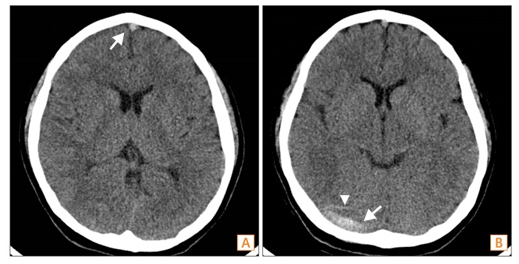
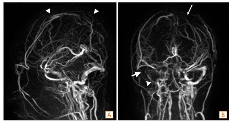
After consultation with neurologists, a cerebrospinal fluid analysis was performed to rule out the possibility of encephalomeningitis. Although testing revealed an elevated intracranial pressure of 220 mmH2O, no white blood cells or red blood cells were detected. Additionally, her protein level was 15.0 mg/dL and her glucose level was 74 mg/dL, further minimizing the likelihood of encephalomeningitis. Therefore, a CT of the brain was performed to rule out other causes of headache and to find the cause of the elevated intracranial pressure. This revealed hyper-attenuations at the anterior and medial superior sagittal sinus, right transverse sinus, and cortical veins, and thus suggested a diagnosis of cerebral venous thrombosis (Fig. 2). The patient then underwent MRI of the brain with venography, resulting in detection of venous thrombosis in the cortical veins, superior sagittal sinus, right transverse sinus, and right internal jugular vein (Fig. 3). Blood tests for homocysteine, factor V, VIII, IX, XI, and vWF antigen were also performed to rule out thrombophilic conditions, and the results were normal. However, her levels of antithrombin III (126%), protein C activity (159%), and protein S activity (131%) were all high. Her D-dimer level (1.4 mg/mL) was also slightly elevated, as may occur during exacerbation of CD.
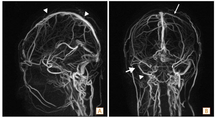
The patient was diagnosed with cerebral venous thrombosis as a complication of CD flare-up, without underlying thrombophilic conditions. She was treated with intravenous heparin (Heparin® 25,000 IU/5mL; JW pharmaceutical, Seoul, Republic of Korea). A follow-up CT scan performed after 7 days of anticoagulation showed resolution of the dense attenuation within the superior sagittal sinus and right transverse sinus (Fig. 4). Her headache and vomiting improved after 9 days of treatment, and completely disappeared after 14 days. An MRI with venography was conducted after 2 weeks of anticoagulation and showed resolution of the venous thrombosis with recanalization of the cortical veins, superior sagittal sinus, and right transverse sinus (Fig. 5).
The patient was discharged on a regimen of rivaroxaban (Xarelto®; Bayer HealthCare AG, Leverkusen, Germany) 10 mg daily, with treatment continued for 6 months. She remained stable, without neurological damage.
IBD can be complicated by a variety of extra-intestinal conditions, including venous thrombosis due to hypercoagulability. In Korea, a few cases of thrombotic complications in patients with CD have been reported (Table 1).56789 In cases of venous thrombosis, cerebral vascular involvement is very rarely observed.45 Cerebral venous thrombosis can be accompanied by neurologic abnormalities caused by rapidly elevating intracranial pressure, and can result in serious adverse events. Therefore, early differential diagnosis of this rare complication is important for CD patients complaining of headache and vomiting. In a study that retrospectively analyzed data from 65 case reports of IBD patients with cerebral venous thrombosis, the average patient age was 29, and women experienced complications more frequently than men (37 vs. 28).3 The incidence of UC was almost double compared with CD (42 vs. 21), and active disease was reported in 78.4% of the cases. The predominant neurological symptoms were headache (80%), seizure (35%), vomiting (29%), hemiplegia or tetraplegia (29%), altered consciousness (21%), vision deficits (12%), and papilledema (7%). The most common site of cerebral venous thrombosis was the superior sagittal sinus (50.7%), and thrombosis in multiple sites occurred in about 50% of cases, like the one we have reported here. The main risk factors for hypercoagulability were anemia (49%) and thrombocytosis (26%); however, there were many cases (23%) accompanied by coagulation abnormalities of fibrinogen, elevated factor VII, elevated factor VIII, protein C deficiency, and protein S deficiency. In our case, cerebral venous thrombosis complicated during flareup of CD, without underlying thrombophilic conditions.
In Korea, only one case has previously documented cerebral venous thrombosis as a complication of CD.5 In that case, a 45-year-old man presented with headaches and was diagnosed with cerebral venous thrombosis as a complication of CD, based on CT and MRI findings. He was treated with heparin and warfarin. However, that patient's CD was stable and not in an active phase, in contrast to our case. In addition, his thrombophilic condition was not fully examined by factor V, VIII, IX, XI, and vWF antigen testing. Therefore, our case report is the first to describe cerebral venous thrombosis during flare-up of CD, without the presence of underlying thrombophilic conditions, in Korea.
Patients with IBD who develop thrombosis often have active disease at the time of thromboembolism.101112 Grainge et al.10 analyzed 13,756 patients with IBD and 71,672 matched controls, and found that 139 patients and 165 controls developed venous thromboembolism. Overall, patients with IBD had a 3.4 times higher risk of developing venous thromboembolism than did controls, with risk increasing up to 8.4 times during a flare-up of IBD. Currently, there is no clear evidence based on randomized controlled trials indicating whether anticoagulation treatment should be used as a preventive measure for patients with IBD. However, anticoagulation therapy for hospitalized patients with active IBD has been associated with lower incidence of venous thrombosis and mortality.1112 Few international guidelines recommended preventive use of anticoagulant therapy for hospitalized patients with active IBD,11 and the Korean IBD treatment guidelines have not addressed this issue.1314
In patients with IBD complicated by cerebral venous thrombosis, favorable outcomes are possible with early diagnosis and appropriate treatment. However, when patients are not treated, the mortality rate can be as high as 50%.3 Therefore, physicians should be especially careful to consider the possibility of cerebral venous thrombosis in patients with IBD, especially when there is active disease, as in our case. Treatment of cerebral venous thrombosis must continue for at least 3 months, but if there are risk factors for thrombosis, coagulation abnormalities, or thrombogenic mutation, then long term treatment of more than 6 months should be maintained.15 Since the underlying IBD can be a cause of thrombosis, treatment of the inflammation itself is essential.11 In our case, the patient's inflammatory activity of CD was rapidly controlled with a biologic agent, and anticoagulation with intravenous heparin reversed her thrombophilic conditions.
Although this is the second case reported in Korea of CD complicated by cerebral venous thrombosis, this is the first case in active CD without underlying thrombophilic conditions. Once again, a high level of concern for cerebral venous thrombosis is important when treating active IBD patients, because anticoagulation may prevent fatal complications.

Financial support: None.

Conflict of interest: None.

  • 1. Bernstein CN, Blanchard JF, Houston DS, Wajda A. The incidence of deep venous thrombosis and pulmonary embolism among patients with inflammatory bowel disease: a population-based cohort study. Thromb Haemost 2001;85:430–434.PMID: 11307809.ArticlePubMedPDF
  • 2. Talbot RW, Heppell J, Dozois RR, Beart RW Jr. Vascular complications of inflammatory bowel disease. Mayo Clin Proc 1986;61:140–145.PMID: 3080643.ArticlePubMed
  • 3. Katsanos AH, Katsanos KH, Kosmidou M, Giannopoulos S, Kyritsis AP, Tsianos EV. Cerebral sinus venous thrombosis in inflammatory bowel diseases. QJM 2013;106:401–413.PMID: 23243293.ArticlePubMedPDF
  • 4. Kwon Y, Koene RJ, Cho Y. A case of cerebral venous sinus thrombosis associated with Crohn's disease: dilemma in management. Gastroenterol Rep (Oxf) [published online ahead of print November 10, 2014]. PMID: 10.1093/gastro/gou079.Article
  • 5. Huh SY, Kim JK, Park SJ. Cerebral venous sinus thrombosis in a patient with Crohn disease. Kosin Med J 2009;24:190–192.
  • 6. Chung C, Kang DH, Kim EA, et al. A case of pulmonary embolism in recurrent ileal Crohn's disease after total colectomy. Korean J Gastrointest Endosc 2001;22:111–115.
  • 7. Chung ES, Kim JH, Jung JH, et al. A case of pulmonary thromboembolism in Crohn's disease. Tuberc Respir Dis 2009;66:370–373.Article
  • 8. Kim W, Kang B, Kim BW, et al. Crohn's disease initially accompanied by deep vein thrombosis and ulnar neuropathy without metronidazole exposure. Gut Liver 2013;7:252–254.PMID: 23560164.ArticlePubMedPMC
  • 9. Oh DY, Jung HK, Kwon KJ, et al. Unusual complication of Crohn's disease: portal hypertension related with rapid progression of portal vein and superior mesenteric vein thrombosis. Korean J Med 2013;84:713–717.Article
  • 10. Grainge MJ, West J, Card TR. Venous thromboembolism during active disease and remission in inflammatory bowel disease: a cohort study. Lancet 2010;375:657–663.PMID: 20149425.ArticlePubMed
  • 11. Papa A, Gerardi V, Marzo M, Felice C, Rapaccini GL, Gasbarrini A. Venous thromboembolism in patients with inflammatory bowel disease: focus on prevention and treatment. World J Gastroenterol 2014;20:3173–3179.PMID: 24695669.ArticlePubMedPMC
  • 12. Zezos P, Kouklakis G, Saibil F. Inflammatory bowel disease and thromboembolism. World J Gastroenterol 2014;20:13863–13878.PMID: 25320522.ArticlePubMedPMC
  • 13. Ye BD, Yang SK, Shin SJ, et al. Guidelines for the management of Crohn's disease. Korean J Gastroenterol 2012;59:141–179.PMID: 22387837.ArticlePubMed
  • 14. Choi CH, Kim YH, Kim YS, et al. Guidelines for the management of ulcerative colitis. Korean J Gastroenterol 2012;59:118–140.PMID: 22387836.ArticlePubMed
  • 15. Einhäupl K, Stam J, Bousser MG, et al. EFNS guideline on the treatment of cerebral venous and sinus thrombosis in adult patients. Eur J Neurol 2010;17:1229–1235.PMID: 20402748.ArticlePubMed
Fig. 1

Endoscopic findings. Endoscopic findings show the longitudinal ulcers (A) A B with cobble stone appearance (B).

ir-14-96-g001.jpg
Fig. 2

Brain CT findings. The CT shows hyperattenuation segments at the anterior and medial superior sagittal sinus (A), right transverse sinus (B, arrow), and cortical veins (B, arrowhead).

ir-14-96-g002.jpg
Fig. 3

Brain MRI with venography. Magnetic resonance venography shows venous thrombosis in the cortical veins (B, thin arrow), superior sagittal sinus (A, arrowheads), right transverse sinus (B, arrow), and right internal jugular vein (B, arrowhead).

ir-14-96-g003.jpg
Fig. 4

Follow-up brain CT after 7 days of anticoagulation. Follow up CT shows resolution of dense attenuation within the superior sagittal sinus (A), right transverse sinus (B, arrow), and cortical veins (B, arrowhead).

ir-14-96-g004.jpg
Fig. 5

Follow-up brain MRI with venography after 2 weeks of anticoagulation. Follow up MRI with venography shows resolved venous thrombosis with recanalization of the cortical veins (B, thin arrow), superior sagittal sinus (A, arrowheads), right transverse sinus (B, arrow), and right internal jugular vein (B, arrowhead).

ir-14-96-g005.jpg
Table 1

Thrombotic Complications in Patients With CD in Korea

ir-14-96-i001.jpg
Reference Sex/age Active disease Type of thrombosis Hypercoagulability Treatment of thrombosis Outcome
Chung et al.6 (2001) F/36 Yes Pulmonary thromboembolism Protein S, Anti thrombin III deficiencies Heparin/warfarin Improved
Chung et al.7 (2009) M/25 Yes Pulmonary thromboembolism No LMWH/warfarin Improved
Huh et al.5 (2009) M/45 No Cerebral venous thrombosis No Heparin/warfarin Improved
Kim et al.8 (2013) M/35 Yes Deep vein thrombosis No Heparin/warfarin Improved
Oh et al.9 (2013) M/42 No Portal vein and superior mesenteric vein thrombosis Protein C, Protein S deficiencies LMWH/warfarin Improved
Current case F/17 Yes Cerebral venous thrombosis No Heparin/rivaroxaban Improved

F, female; M, male; LMWH, low molecular weight heparin.

Figure & Data

REFERENCES

    Citations

    Citations to this article as recorded by  
    • Cerebral venous thrombosis during relapse of ulcerative colitis: Case report with review of literature
      S. Bouchal, B. Alami, N. Chtaou, M. Abkari, M. Maaroufi, F. Belahsen
      JMV-Journal de Médecine Vasculaire.2021; 46(1): 22.     CrossRef
    • Direct oral anticoagulants in treatment of cerebral venous thrombosis: a systematic review
      Gauruv Bose, Justin Graveline, Vignan Yogendrakumar, Risa Shorr, Dean A Fergusson, Gregoire Le Gal, Jonathan Coutinho, Marcelo Mendonça, Miguel Viana-Baptista, Simon Nagel, Dar Dowlatshahi
      BMJ Open.2021; 11(2): e040212.     CrossRef
    • The Twists and Turns of Diagnosis and Treatment of Pediatric Neuro-Behcet's Disease: A Case Report and Literature Review
      Qiao Zhang, Yizhen Luo, Jianli Zhou, Shaoming Zhou, Zhaoxia Wang
      Frontiers in Pediatrics.2021;[Epub]     CrossRef
    • Recurrent Unprovoked Venous Thrombosis (Cerebral Sinus Thrombosis and Mesenteric Vein Thrombosis) in Young Patient with Crohn’s Disease: A Case Report and Review
      Abdullah Mohammed Albishi, Rafaat Chakik, Ali Saleh Alshamrani
      Case Reports in Gastrointestinal Medicine.2020; 2020: 1.     CrossRef
    • Use of direct oral anticoagulants in cerebral venous thrombosis: a systematic review
      Sen Sheng, Krishina Nalleballe, Naga V. Pothineni, Rohan Sharma, Aliza Brown, Hisham Elkhider, Saritha Ranabothu, Nidhi Kapoor, Kelly-Ann Patrice, Sanjeeva Onteddu
      Blood Coagulation & Fibrinolysis.2020; 31(8): 501.     CrossRef
    • Direct oral anticoagulant use in patients with thrombophilia, antiphospholipid syndrome or venous thrombosis of unusual sites: A narrative review
      Laurent Bertoletti, Ygal Benhamou, Yannick Béjot, Sylvestre Marechaux, Saida Cheggour, Boris Aleil, Nicolas Lellouche, Jean-Guillaume Dillinger, Aurélien Delluc
      Blood Reviews.2018; 32(4): 272.     CrossRef
    • A case of ulcerative colitis presenting with cerebral venous thrombosis
      Junghwan Lee, Sung Wook Hwang, Jinhee Lee, Kyung Hwa Jung, Ha Il Kim, Sang Hyoung Park, Dong-Hoon Yang, Byong Duk Ye, Jeong-Sik Byeon, Seung-Jae Myung, Suk-Kyun Yang
      Intestinal Research.2018; 16(2): 306.     CrossRef
    • Rivaroxaban and Apixaban for Initial Treatment of Acute Venous Thromboembolism of Atypical Location
      Dawid T. Janczak, Malgorzata K. Mimier, Robert D. McBane, Patrick S. Kamath, Benjamin S. Simmons, Dalene M. Bott-Kitslaar, Charles J. Lenz, Emily R. Vargas, David O. Hodge, Waldemar E. Wysokinski
      Mayo Clinic Proceedings.2018; 93(1): 40.     CrossRef
    • Use of Novel Oral Anticoagulant to Treat Pulmonary Thromboembolism in Patient with Ulcerative Colitis Superinfected Cytomegalovirus Colitis
      Seok-Hwan Kim, Sunhee Jang, Yegyu Sung, Jun Kyu Park, Yunjung Park, Jintak Yun, Sang-Bum Kang
      The Korean Journal of Gastroenterology.2017; 70(1): 44.     CrossRef

    • PubReader PubReader
    • ePub LinkePub Link
    • Cite
      CITE
      export Copy Download
      Close
      Download Citation
      Download a citation file in RIS format that can be imported by all major citation management software, including EndNote, ProCite, RefWorks, and Reference Manager.

      Format:
      • RIS — For EndNote, ProCite, RefWorks, and most other reference management software
      • BibTeX — For JabRef, BibDesk, and other BibTeX-specific software
      Include:
      • Citation for the content below
      Cerebral venous thrombosis in a patient with Crohn's disease
      Intest Res. 2016;14(1):96-101.   Published online January 26, 2016
      Close
    • XML DownloadXML Download
    Figure
    • 0
    • 1
    • 2
    • 3
    • 4
    Related articles
    Cerebral venous thrombosis in a patient with Crohn's disease
    Image Image Image Image Image
    Fig. 1 Endoscopic findings. Endoscopic findings show the longitudinal ulcers (A) A B with cobble stone appearance (B).
    Fig. 2 Brain CT findings. The CT shows hyperattenuation segments at the anterior and medial superior sagittal sinus (A), right transverse sinus (B, arrow), and cortical veins (B, arrowhead).
    Fig. 3 Brain MRI with venography. Magnetic resonance venography shows venous thrombosis in the cortical veins (B, thin arrow), superior sagittal sinus (A, arrowheads), right transverse sinus (B, arrow), and right internal jugular vein (B, arrowhead).
    Fig. 4 Follow-up brain CT after 7 days of anticoagulation. Follow up CT shows resolution of dense attenuation within the superior sagittal sinus (A), right transverse sinus (B, arrow), and cortical veins (B, arrowhead).
    Fig. 5 Follow-up brain MRI with venography after 2 weeks of anticoagulation. Follow up MRI with venography shows resolved venous thrombosis with recanalization of the cortical veins (B, thin arrow), superior sagittal sinus (A, arrowheads), right transverse sinus (B, arrow), and right internal jugular vein (B, arrowhead).
    Cerebral venous thrombosis in a patient with Crohn's disease

    Thrombotic Complications in Patients With CD in Korea

    ReferenceSex/ageActive diseaseType of thrombosisHypercoagulabilityTreatment of thrombosisOutcome
    Chung et al.6 (2001)F/36YesPulmonary thromboembolismProtein S, Anti thrombin III deficienciesHeparin/warfarinImproved
    Chung et al.7 (2009)M/25YesPulmonary thromboembolismNoLMWH/warfarinImproved
    Huh et al.5 (2009)M/45NoCerebral venous thrombosisNoHeparin/warfarinImproved
    Kim et al.8 (2013)M/35YesDeep vein thrombosisNoHeparin/warfarinImproved
    Oh et al.9 (2013)M/42NoPortal vein and superior mesenteric vein thrombosisProtein C, Protein S deficienciesLMWH/warfarinImproved
    Current caseF/17YesCerebral venous thrombosisNoHeparin/rivaroxabanImproved

    F, female; M, male; LMWH, low molecular weight heparin.

    Table 1 Thrombotic Complications in Patients With CD in Korea

    F, female; M, male; LMWH, low molecular weight heparin.


    Intest Res : Intestinal Research
    Close layer
    TOP