Intest Res Search

CLOSE


Intest Res > Volume 21(4); 2023 > Article
Singh, Midha, Narang, Kedia, Mahajan, Dhoble, Kahlon, Dhaliwal, Tripathi, Kalra, Jain, Bansal, Banerjee, Desai, Dutta, Ahuja, and Sood: Low prevalence of primary sclerosing cholangitis in patients with inflammatory bowel disease in India

Abstract

Background/Aims

Primary sclerosing cholangitis (PSC) represents the most common hepatobiliary extraintestinal manifestation of inflammatory bowel disease (IBD), including ulcerative colitis (UC) and Crohn’s disease (CD). Limited data exist on PSC in patients with IBD from India. We aimed to assess the prevalence and disease spectrum of PSC in Indian patients with IBD.

Methods

Database of IBD patients at 5 tertiary care IBD centers in India were analyzed retrospectively. Data were extracted and the prevalence of PSC-IBD was calculated.

Results

Forty-eight patients out of 12,216 patients with IBD (9,231 UC, 2,939 CD, and 46 IBD unclassified) were identified to have PSC, resulting in a prevalence of 0.39%. The UC to CD ratio was 7:1. Male sex and pancolitis (UC) or colonic CD were more commonly associated with PSC-IBD. The diagnosis of IBD preceded the diagnosis of PSC in most of the patients. Majority of the patients were symptomatic for liver disease at diagnosis. Eight patients (16.66%) developed cirrhosis, 5 patients (10.41%), all UC, developed malignancies (3 colorectal cancer [6.25%] and 2 cholangiocarcinoma [4.16%]), and 3 patients died (2 decompensated liver disease [4.16%] and 1 cholangiocarcinoma [2.08%]) on follow-up. None of the patients mandated surgical therapy for IBD.

Conclusions

Concomitant PSC in patients with IBD is uncommon in India and is associated with lower rates of development of malignancies.

INTRODUCTION

Primary sclerosing cholangitis (PSC), a chronic progressive inflammatory disease of the intrahepatic and/or extrahepatic bile ducts, represents the main hepatobiliary extraintestinal manifestation of inflammatory bowel disease (IBD) [1,2]. The prevalence of PSC in IBD (PSC-IBD) varies between 3% to 8% for patients with ulcerative colitis (UC) and 2% to 3% for patients with Crohn’s disease (CD), while PSC is associated with IBD in 50% to 90% patients [3,4]. Though the exact pathogenesis of both IBD and PSC are not known, the 2 are believed to share a common pathophysiology, influenced by immune-mediated processes (gut lymphocyte homing), immunogenic susceptibility, increased intestinal permeability, dysbiotic gut microbiota, genetic predisposition, disorders of biliary epithelial cells and altered bile acid metabolism [5-9]. The PSC-IBD has been described as a unique entity, different from IBD, characterized by an increased incidence of pancolitis, backwash ileitis, and rectal sparing; though the colitis tends to be clinically and often endoscopically quiescent [10-13]. An increased risk of colorectal cancer (CRC) in patients with PSC and IBD drives the differences in approach to management of PSC with IBD as compared to IBD alone, the former including lifelong annual surveillance colonoscopy [14-17].
Though the association between PSC and IBD is well recognized, there remains uncertainty on the magnitude of the problem. A recent systematic review and meta-analysis estimated the global prevalence of PSC in patients with IBD at 2.16%, with appreciable regional variations [18]. Limited data exist on the prevalence and disease phenotype of PSC with IBD from India [19,20]. This multicenter study aimed to investigate the prevalence of PSC with IBD, describe the disease phenotype (clinical features, disease characteristics, and disease-related complications) and evaluate regional variations in the spectrum of PSC in patients with IBD in India.

METHODS

1. Study Design

This study is a retrospective analysis of prospectively maintained IBD databases at 5 tertiary care centers across India: Dayanand Medical College and Hospital, Ludhiana (north India); All India Institute of Medical Sciences, New Delhi (north India); Postgraduate Institute of Medical Education and Research, Chandigarh (north India); P.D. Hinduja National Hospital and Medical Research Centre, Mumbai (west India); and Asian Institute of Gastroenterology, Hyderabad (south India). The respective databases were reviewed for a period between January 1991 and December 2020. The study was approved by the Ethics Committee of Dayanand Medical College and Hospital (IRB No. DMCH/R&D/2020/23). This study is a retrospective study and so informed consent was waived. A proportion of the patients reported in the current study were also included in a previous single-center study describing the prevalence and spectrum of extraintestinal manifestations in patients with IBD [19].

2. Study Population

Patients with established diagnosis of IBD (based on the European Crohn’s and Colitis Organisation and European Society of Gastrointestinal and Abdominal Radiology Guidelines) who were found to have concomitant PSC (diagnosed on the basis of biochemical cholestasis [elevated alkaline phosphatase] and cholangiographic evidence of strictures/ectasias of intrahepatic and/or extrahepatic bile ducts) were included for analysis [21,22].
The details on demographic profile (including age at diagnosis of PSC-IBD and sex), clinical history (disease diagnosis, clinical presentation, disease extent, severity, and behavior), complications including cirrhosis, CRC and cholangiocarcinoma (CCA) and medical/surgical treatment were extracted from the databases. Montreal classification was used to assess the disease phenotype in adults with IBD. Disease severity was defined by the Mayo Clinic score in patients with UC and Harvey Bradshaw Index in patients with CD.
Patients with missing/incomplete data, follow-up < 6 months and a secondary cause for sclerosing cholangitis (such as infectious or ischemic cholangiopathy, IgG4 associated cholangitis, portal cavernoma cholangiopathy, drug-induced liver injury, choledocholithiasis, and surgical trauma to biliary tract) were excluded.

3. Statistical Analysis

Quantitative data were presented as mean ± standard deviation or median and interquartile range (IQR). Categorical data were summarized as the percentage of the group total. Categorical data were compared using either the chi-square test (for parametric data) or the Kruskal-Wallis one-way analysis of variance test (for non-parametric data). Fischer exact test was performed when the anticipated frequency was less than 5. A probability value (P-value) less than 0.05 was considered statistically significant. All statistical calculations were done using SPSS version 21 (IBM Corp., Armonk, NY, USA) statistical program for Microsoft Windows.

RESULTS

1. Prevalence of PSC in Patients with IBD

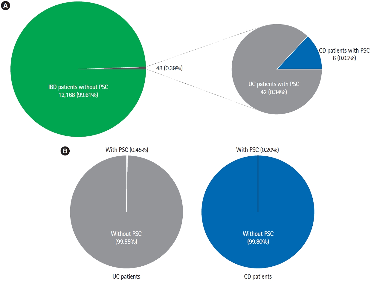
Records of 12,216 patients with IBD (9,231 UC, 2,939 CD, and 46 IBD unclassified) were analyzed. Forty-eight patients were identified to have PSC, resulting in a prevalence of 0.39%. Of the 48 patients diagnosed with PSC, 42 (87.50%) had UC while 6 (12.50%) had CD. The cumulative prevalence of PSC was 0.45% in patients with UC and 0.20% in patients with CD (Fig. 1). The mean Mayo Clinic score and Harvey Bradshaw Index were 5.26 ± 2.41 and 5.17 ± 1.33 in patients with UC-PSC and CD-PSC, respectively.

2. Characteristics of the IBD Patients Diagnosed with PSC

Majority of the patients (n = 27, 56.25%) were males. In nearly 70% of the patients, the diagnosis of IBD preceded the diagnosis of PSC with a mean duration of 43.25 ± 74.66 months between the diagnoses of the 2 entities. In only 4 patients (8.33%), PSC was diagnosed before the diagnosis of IBD was established. Approximately two-thirds (69.05%) of the patients with UC-PSC had pancolitis, while isolated colonic involvement (50%) followed by ileo-colonic disease (33.33%) was the most common disease location in patients with CD-PSC. The frequency of stricturing and/or penetrating disease was low (16.66%) in patients with CD-PSC. The clinical characteristics of the patients with PSC and IBD are summarized in Table 1.

3. Clinical Presentation and Complications of PSC in Patients with IBD

A majority of the patients (n = 32, 68.75%) were symptomatic for liver disease at the time of diagnosis of PSC-IBD. The common presenting symptoms were fatigue (n = 31, 64.58%), abdominal pain (n = 14, 29.16%), pruritus (n = 12, 25%), and jaundice (n = 7, 14.58%). One patient had cirrhosis at the time of diagnosis of PSC (Table 2). The median alkaline phosphatase at the time of diagnosis was 417.0 IU/L (IQR, 293.5-637.0 IU/L). All the patients had evidence of cholangiographic changes of beading/stricturing and/or ectasias.
The patients were followed up for a median of 66 months (IQR, 27-129 months). During the follow-up period, 7 more patients developed cirrhosis. Five patients (10.41%), all UC, developed malignancies (3 CRC [6.25%] and 2 CCA [4.16%]) (Fig. 2). Three patients died during follow-up (2 decompensated liver disease and 1 CCA) (Table 2).
All the patients were on concomitant therapy with ursodeoxycholic acid for PSC. Eight patients with dominant biliary strictures required endoscopic balloon dilatation. Two patients needed liver transplant for decompensated liver disease, while another 2 underwent colectomy with adjuvant chemotherapy for CRC. Three patients (2 CCA and 1 CRC) received palliative chemotherapy for associated malignancy. None of the patients mandated surgical therapy for IBD (Table 2).

DISCUSSION

This multicenter study evaluated the relationship and characteristics of IBD and PSC in a large cohort of IBD patients from India. The prevalence of PSC in patients with IBD was 0.39%. The prevalence of PSC in UC and CD separately was 0.45% and 0.20%. The prevalence rate in the current study is lower than the reported prevalence in previous studies from Westernized countries [18,23,24]. However the PSC prevalence rate in IBD is similar to previously reported rates of 0.3% in India and 0.48% in the Inflammatory Bowel Disease-Emerging Nations’ Consortium [19,20]. PSC (with or without concomitant IBD) exhibits wide variations in its geo-epidemiology, with lower prevalence rates reported from Asia as compared to North America and Europe. The prevalence rates of PSC-IBD in the current study are amongst the lowest, even in Asia [18]. This could be attributed to heterogeneity in genetic, ethnic, environmental, and other risk factors that lead to development of PSC, suggesting that though the disease phenotypes maybe similar, demographic differences exist in IBD among Asian and Western countries [25].
Majority of the patients with PSC-IBD (both UC and CD) concurrence were males (56.25%) and had their IBD diagnosed before PSC (70%). The ratio of UC-PSC and CD-PSC concurrence in the current study was 7:1, indicating higher prevalence of PSC in patients with UC as compared to CD. Consistent with the previous reports, pancolitis and colonic disease location were the commonest disease extent and location in patients with UC and CD respectively, suggesting colonic involvement as the hallmark of the PSC-IBD phenotype. However, in contrast to the earlier studies, where none of the patients with proctosigmoiditis had PSC, 30% of UC-PSC patients in the current study had proctosigmoiditis (disease extent E2) [26-28]. Whether this represents a distinct phenotype of PSC-IBD that behaves differently form the pancolitis associated PSC-IBD is not known and needs to be evaluated. Only 6.25% of patients with PSC-IBD were current or former smokers. This is congruous with decreased risk of PSC among smokers described earlier [29].
Most of the patients with PSC-IBD were symptomatic at diagnosis. This is in contrast to the earlier descriptions of PSC being asymptomatic and detected on screening for liver disease in patients with IBD [30]. A higher proportion of patients being symptomatic in the current study could either imply late diagnosis of PSC due to challenges in adhering to PSC screening protocols [31] or a different spectrum of PSC-IBD in India. Although statistically insignificant, a greater proportion of patients with CD-PSC developed cirrhosis. On the contrary, malignancies (either CRC or CCA) developed more frequently in patients with UC-PSC. The rates of liver transplantation and death were lower in patients with CD-PSC. Though evaluating the effect of IBD type on the clinical presentation and complications of PSC-IBD was not the objective of the current study, our findings suggest a natural history of CD-PSC distinct from the UC-PSC. Similar findings have been reported previously, where patients with CD-PSC progressed less commonly to cancer, liver transplantation, or death [32,33].
Approximately 10% of the patients with PSC-IBD progressed to develop malignancies. The prevalence of CRC and CCA in PSC-IBD was 6.25% and 4.17%, respectively. CCA has been reported to develop in up to 5%-20% of patients with PSC [34,35]. The prevalence of CRC in patients with IBD varies between 2%-3% in India and increases substantially in the second and third decade after the onset of IBD even though the incidence of sporadic CRC is low [36-38]. This paradox suggests that inflammation-driven CRC has similar incidence rates in the East as well as the West. Though the prevalence of CRC in PSC-IBD is twice the prevalence in UC without PSC, the rates are much lower than the 20% to 30% prevalence rates described previously [39,40]. The lower rates of CRC can be attributed to distinct genetic and gut microbial composition of Indians as compared to the Caucasians, apart from variations in the Farnesoid X receptor and bile acid metabolic pathways, which play an integral role in the development of malignancies in patients with PSC-IBD [41-43]. The lower incidence of CRC in the present cohort could also possibly be related to a shorter follow-up period and low annual CRC screening rates. More prospective longitudinal studies are needed to better determine the natural history and formulate cost-effective population specific policies for surveillance of cancers in PSC-IBD patients.
This study is the first nationwide study determining the prevalence of PSC in patients with IBD from India. The strengths of our study include large sample size and strict criteria of a combination of biochemical cholestasis and positive cholangiography for diagnosis of PSC. However, retrospective nature of the study may have resulted in an underestimation of the true prevalence of PSC-IBD as patients with missing data were excluded. Patients with PSC who do not have clinical symptoms of IBD may have endoscopic and/or histological features consistent with IBD. As patients with PSC who did not have intestinal symptoms were not evaluated in the current study, underreporting of the prevalence of PSC-IBD is possible, though the proportion of such patients is expected to be small. Data on liver biopsy was not available and therefore patients with small duct PSC or patients with normal biochemistry could have been missed. Additionally, the details on proportion of patients with backwash ileitis and rectal sparing, reportedly more prevalent in patients with PSC-IBD, were not available.
In conclusion, this multicenter nationwide study demonstrates low prevalence rates (0.39%) of PSC in patients with IBD in India. Males, patients with UC and patients with pancolitis (UC) or colonic CD were more likely to receive a diagnosis of concomitant PSC and IBD. Majority of the patients were symptomatic of PSC at the time of diagnosis. The prevalence of CRC and CCA was low as compared to other reports from Asia and the West. The data presented in the current study provides a blueprint of the spectrum of PSC-IBD in India, which is disparate from that reported in the literature. More data is needed to describe the true spectrum and long-term outcome of PSC-IBD in India. To achieve this, it is important to recognize and carefully screen all IBD patients for PSC, as an early appropriate diagnosis is imperative to prevent complications.

ADDITIONAL INFORMATION

Funding Source

The authors received no financial support for the research, authorship, and/or publication of this article.

Conflict of Interest

Sood A is an editorial board member of the journal but was not involved in the peer reviewer selection, evaluation, or decision process of this article. No other potential conflicts of interest relevant to this article were reported.

Data Availability Statement

The data that support the findings of this study are available from the corresponding author upon reasonable request.

Author Contributions

Conceptualization: Midha V, Sood A. Data curation: Singh A, Narang V, Kedia S, Dhoble P, Kahlon BK, Dhaliwal AS, Tripathi A, Kalra S, Bansal N, Banerjee R, Desai D, Dutta U, Ahuja V. Formal analysis: Singh A, Narang V, Bansal N. Investigation: Singh A, Midha V, Sood A. Methodology: Singh A, Midha V, Sood A. Project administration: Midha V, Sood A. Resources: Singh A, Midha V, Narang V, Dhoble P, Jain NP, Desai D, Sood A. Software: Singh A, Kahlon BK, Bansal N. Supervision: Midha V, Sood A. Visualization: Singh A, Kedia S, Mahajan R, Jain NP, Bansal N, Desai D, Ahuja V, Sood A. Writing - original draft: Singh A, Narang V. Writing - review & editing: all authors. Approval of final manuscript: all authors.

Fig. 1.
PSC in patients with IBD. (A) Prevalence and distribution of PSC in patients with IBD. (B) The prevalence of PSC in patients with UC and CD. PSC, primary sclerosing cholangitis; IBD, inflammatory bowel disease; UC, ulcerative colitis; CD, Crohn’s disease.
ir-2022-00087f1.jpg
Fig. 2.
Prevalence of malignancies in patients with primary sclerosing cholangitis (PSC) and inflammatory bowel disease (IBD).
ir-2022-00087f2.jpg
Table 1.
Characteristics of Patients with PSC with IBD
Characteristics Value (n = 48)
Male sex 27 (56.25)
Age at diagnosis of IBD (yr) 33.67 ± 12.16
Age at diagnosis of PSC (yr) 37.27 ± 12.78
Follow up (mo) 66 (27-129)
Current or former smoker 3 (6.25)
PSC diagnosed before IBD 4 (8.33)
IBD diagnosed before PSC 34 (70.83)
IBD and PSC diagnosed simultaneously 10 (20.83)
Interval between diagnosis of IBD and PSC (mo) 43.25 ± 74.66
Type of IBD
 Ulcerative colitis 42 (87.5)
 Crohn’s disease 6 (12.5)
Disease extent (ulcerative colitis)
 Proctitis -
 Left sided colitis 13 (30.95)
 Pancolitis 29 (69.05)
Mayo Clinic score at diagnosis of IBD 5.26 ± 2.41
Endoscopic Mayo Clinic score at diagnosis of IBD 1.64 ± 0.62
Disease classification (Crohn’s disease)
 Age at diagnosis
  < 17 yr -
  17-40 yr 4 (66.66)
  > 40 yr 2 (33.33)
 Disease location
  Ileal 1 (16.66)
  Colonic 3 (50.00)
  Ileo-colonic 2 (33.33)
 Disease behavior
  Non-stricturing, non-penetrating 5 (83.33)
  Stricturing 1 (16.66)
 Penetrating -
Perianal disease Nil
Harvey Bradshaw Index at diagnosis of IBD 5.17 ± 1.33
Patients with other extraintestinal manifestationsa 9 (18.75)
 Arthritis 5 (10.41)
 Ocular 4 (8.33)
 Gall stones 1 (2.08)
 Erythema nodosum 1 (2.08)
Family history of IBD 1 (2.08)
Previous appendectomy 4 (8.33)
Previous exposure to anti-tubercular therapy 7 (14.58)
Treatment for IBD
 5-Aminosalicylates 39 (81.25)
 Thiopurines 14 (29.16)
 Anti-TNF 1 (2.08)
 Corticosteroids 20 (41.66)

Values are presented as number (%), mean±standard deviation, or median (interquartile range).

a More than one extraintestinal manifestations were present in all these patients.

PSC, primary sclerosing cholangitis; IBD, inflammatory bowel disease; TNF, tumor necrosis factor.

Table 2.
Clinical Presentation and Complications of PSC in Patients with Inflammatory Bowel Disease
Variable Ulcerative colitis (n = 42) Crohn’s disease (n = 6) Total (n = 48)
Clinical presentation of PSC
 Asymptomatic with abnormal liver function tests 13 (30.95) 2 (33.33) 15 (31.25)
 Symptomatic with abnormal liver function tests 29 (69.04) 4 (66.66) 33 (68.75)
Complications
 Cirrhosis 6 (14.28) 2 (33.33) 8 (16.66)
 CRC 3 (7.14) - 3 (6.25)
 CCA 2 (4.76) - 2 (4.16)
 Death 3 (7.14) - 3 (6.25)
  Decompensated liver disease 2 (4.76) - 2 (4.16)
  CCA 1 (2.38) - 1 (2.08)
Treatment of complications
 Liver transplant for decompensated cirrhosis 2 (4.76) - 2 (4.16)
 Colectomy with adjuvant chemotherapy (CRC) 2 (4.76) - 2 (4.16)
 Palliative chemotherapy (CRC) 1 (2.38) - 1 (2.08)
 Palliative chemotherapy (CCA) 2 (4.76) - 2 (4.16)
Treatment received for PSC
 Ursodeoxycholic acid 42 (100) 6 (100) 48 (100)
 Endoscopic balloon dilatation for dominant stricture 7 (16.66) 1 (16.66) 8 (16.66)

Values are presented as number (%).

PSC, primary sclerosing cholangitis; CRC, colorectal cancer; CCA, cholangiocarcinoma.

REFERENCES

1. Smith MP, Loe RH. Sclerosing cholangitis; review of recent case reports and associated diseases and four new cases. Am J Surg 1965;110:239-246.
pmid
2. Lazaridis KN, LaRusso NF. Primary sclerosing cholangitis. N Engl J Med 2016;375:1161-1170.
crossref pmid pmc
3. de Vries AB, Janse M, Blokzijl H, Weersma RK. Distinctive inflammatory bowel disease phenotype in primary sclerosing cholangitis. World J Gastroenterol 2015;21:1956-1971.
crossref pmid pmc
4. Heikius B, Niemelä S, Lehtola J, Karttunen T, Lähde S. Hepatobiliary and coexisting pancreatic duct abnormalities in patients with inflammatory bowel disease. Scand J Gastroenterol 1997;32:153-161.
crossref pmid
5. Duboc H, Rajca S, Rainteau D, et al. Connecting dysbiosis, bileacid dysmetabolism and gut inflammation in inflammatory bowel diseases. Gut 2013;62:531-539.
crossref pmid
6. Quraishi MN, Sergeant M, Kay G, et al. The gut-adherent microbiota of PSC-IBD is distinct to that of IBD. Gut 2017;66:386-388.
crossref
7. Grant AJ, Lalor PF, Salmi M, Jalkanen S, Adams DH. Homing of mucosal lymphocytes to the liver in the pathogenesis of hepatic complications of inflammatory bowel disease. Lancet 2002;359:150-157.
crossref pmid
8. Jostins L, Ripke S, Weersma RK, et al. Host-microbe interactions have shaped the genetic architecture of inflammatory bowel disease. Nature 2012;491:119-124.
pmid pmc
9. Weismüller TJ, Wedemeyer J, Kubicka S, Strassburg CP, Manns MP. The challenges in primary sclerosing cholangitis: aetiopathogenesis, autoimmunity, management and malignancy. J Hepatol 2008;48 Suppl 1:S38-S57.
pmid
10. Loftus EV Jr, Harewood GC, Loftus CG, et al. PSC-IBD: a unique form of inflammatory bowel disease associated with primary sclerosing cholangitis. Gut 2005;54:91-96.
crossref pmid pmc
11. Sinakos E, Samuel S, Enders F, Loftus EV Jr, Sandborn WJ, Lindor KD. Inflammatory bowel disease in primary sclerosing cholangitis: a robust yet changing relationship. Inflamm Bowel Dis 2013;19:1004-1009.
pmid
12. Palmela C, Peerani F, Castaneda D, Torres J, Itzkowitz SH. Inflammatory bowel disease and primary sclerosing cholangitis: a review of the phenotype and associated specific features. Gut Liver 2018;12:17-29.
crossref pmid pmc
13. Beheshti-Maal A, Tamimi A, Iravani S, et al. PSC associated inflammatory bowel disease: a distinct entity. Expert Rev Gastroenterol Hepatol 2022;16:129-139.
crossref pmid
14. Lindström L, Lapidus A, Ost A, Bergquist A. Increased risk of colorectal cancer and dysplasia in patients with Crohn’s colitis and primary sclerosing cholangitis. Dis Colon Rectum 2011;54:1392-1397.
crossref pmid
15. Claessen MM, Lutgens MW, van Buuren HR, et al. More rightsided IBD-associated colorectal cancer in patients with primary sclerosing cholangitis. Inflamm Bowel Dis 2009;15:1331-1336.
crossref pmid
16. Kornfeld D, Ekbom A, Ihre T. Is there an excess risk for colorectal cancer in patients with ulcerative colitis and concomitant primary sclerosing cholangitis? A population based study. Gut 1997;41:522-525.
crossref pmid pmc
17. Wijnands AM, de Jong ME, Lutgens MW, et al. Prognostic factors for advanced colorectal neoplasia in inflammatory bowel disease: systematic review and meta-analysis. Gastroenterology 2021;160:1584-1598.
crossref pmid
18. Barberio B, Massimi D, Cazzagon N, Zingone F, Ford AC, Savarino EV. Prevalence of primary sclerosing cholangitis in patients with inflammatory bowel disease: a systematic review and meta-analysis. Gastroenterology 2021;161:1865-1877.
crossref pmid
19. Singh B, Kedia S, Konijeti G, et al. Extraintestinal manifestations of inflammatory bowel disease and intestinal tuberculosis: frequency and relation with disease phenotype. Indian J Gastroenterol 2015;34:43-50.
crossref pmid pdf
20. Banerjee R, Pal P, Hilmi I, et al. Emerging inflammatory bowel disease demographics, phenotype, and treatment in South Asia, South-East Asia, and Middle East: preliminary findings from the inflammatory bowel disease-emerging nations’ consortium. J Gastroenterol Hepatol 2022;37:1004-1015.
crossref pmid pdf
21. Maaser C, Sturm A, Vavricka SR, et al. ECCO-ESGAR guideline for diagnostic assessment in IBD part 1: initial diagnosis, monitoring of known IBD, detection of complications. J Crohns Colitis 2019;13:144-164.
crossref pmid
22. Chapman MH, Thorburn D, Hirschfield GM, et al. British Society of Gastroenterology and UK-PSC guidelines for the diagnosis and management of primary sclerosing cholangitis. Gut 2019;68:1356-1378.
crossref pmid pmc
23. Guerra I, Bujanda L, Castro J, et al. Clinical characteristics, associated malignancies and management of primary sclerosing cholangitis in inflammatory bowel disease patients: a multicentre retrospective cohort study. J Crohns Colitis 2019;13:1492-1500.
pmid
24. Lunder AK, Hov JR, Borthne A, et al. Prevalence of sclerosing cholangitis detected by magnetic resonance cholangiography in patients with long-term inflammatory bowel disease. Gastroenterology 2016;151:660-669.e4.
crossref pmid
25. Banerjee R, Pal P, Nugent Z, et al. IBD in India: similar phenotype but different demographics than the west. J Clin Gastroenterol 2020;54:725-732.
pmid
26. Olsson R, Danielsson A, Järnerot G, et al. Prevalence of primary sclerosing cholangitis in patients with ulcerative colitis. Gastroenterology 1991;100(5 Pt 1):1319-1323.
crossref pmid
27. Wewer V, Gluud C, Schlichting P, Burcharth F, Binder V. Prevalence of hepatobiliary dysfunction in a regional group of patients with chronic inflammatory bowel disease. Scand J Gastroenterol 1991;26:97-102.
crossref pmid
28. Roberts H, Rai SN, Pan J, et al. Extraintestinal manifestations of inflammatory bowel disease and the influence of smoking. Digestion 2014;90:122-129.
crossref pmid pdf
29. Wijarnpreecha K, Panjawatanan P, Mousa OY, Cheungpasitporn W, Pungpapong S, Ungprasert P. Association between smoking and risk of primary sclerosing cholangitis: a systematic review and meta-analysis. United European Gastroenterol J 2018;6:500-508.
crossref pmid pmc pdf
30. Kaplan GG, Laupland KB, Butzner D, Urbanski SJ, Lee SS. The burden of large and small duct primary sclerosing cholangitis in adults and children: a population-based analysis. Am J Gastroenterol 2007;102:1042-1049.
crossref pmid
31. Banerjee R, Pal P, Mak JW, Ng SC. Challenges in the diagnosis and management of inflammatory bowel disease in resourcelimited settings in Asia. Lancet Gastroenterol Hepatol 2020;5:1076-1088.
crossref pmid
32. Halliday JS, Djordjevic J, Lust M, et al. A unique clinical phenotype of primary sclerosing cholangitis associated with Crohn’s disease. J Crohns Colitis 2012;6:174-181.
crossref pmid
33. Navaneethan U, Venkatesh PG, Jegadeesan R, et al. Comparison of outcomes for patients with primary sclerosing cholangitis associated with ulcerative colitis and Crohn’s disease. Gastroenterol Rep (Oxf) 2016;4:43-49.
crossref pmid
34. Razumilava N, Gores GJ. Cholangiocarcinoma. Lancet 2014;383:2168-2179.
crossref pmid pmc
35. Björnsson E, Angulo P. Cholangiocarcinoma in young individuals with and without primary sclerosing cholangitis. Am J Gastroenterol 2007;102:1677-1682.
crossref pmid
36. Bopanna S, Kedia S, Das P, et al. Long-term follow-up reveals high incidence of colorectal cancer in Indian patients with inflammatory bowel disease. United European Gastroenterol J 2017;5:708-714.
crossref pmid pmc pdf
37. Desai D, Shah S, Deshmukh A, et al. Colorectal cancers in ulcerative colitis from a low-prevalence area for colon cancer. World J Gastroenterol 2015;21:3644-3649.
crossref pmid pmc
38. Bopanna S, Roy M, Das P, et al. Role of random biopsies in surveillance of dysplasia in ulcerative colitis patients with high risk of colorectal cancer. Intest Res 2016;14:264-269.
crossref pmid pmc
39. Karlsen TH. Primary sclerosing cholangitis: 50 years of a gutliver relationship and still no love? Gut 2016;65:1579-1581.
crossref pmid
40. Ananthakrishnan AN, Cagan A, Gainer VS, et al. Mortality and extraintestinal cancers in patients with primary sclerosing cholangitis and inflammatory bowel disease. J Crohns Colitis 2014;8:956-963.
crossref pmid pmc
41. Hill MJ. Bile flow and colon cancer. Mutat Res 1990;238:313-320.
crossref pmid
42. Farhana L, Nangia-Makker P, Arbit E, et al. Bile acid: a potential inducer of colon cancer stem cells. Stem Cell Res Ther 2016;7:181.
crossref pmid pmc pdf
43. Garrett WS. The gut microbiota and colon cancer. Science 2019;364:1133-1135.
crossref pmid
TOOLS
Share :
Facebook Twitter Linked In Google+ Line it
METRICS Graph View
  • 3 Web of Science
  • 3 Crossref
  • 3 Scopus
  • 3,228 View
  • 542 Download
We recommend


ABOUT
ARTICLE & TOPICS
Article Category

Browse all articles >

TOPICS

Browse all articles >

BROWSE ARTICLES
EDITORIAL POLICY
AUTHOR INFORMATION
Editorial Office
Room 310, Lotte Gold Rose II, 31 Seolleung-ro 86-gil, Gangnam-gu, Seoul 06193, Korea
Tel: +82-2-957-6145    Fax: +82-2-957-6146    E-mail: thekasid@irjournal.org                

Copyright © 2024 by Korean Association for the Study of Intestinal Diseases.

Developed in M2PI

Close layer
prev next